IMAGE OF THE WEEK 2014
IMAGE 5
HEART VALVES REPLACEMENT: PART 2 -
MITRAL VALVE STENOSIS
|
 |
|
|
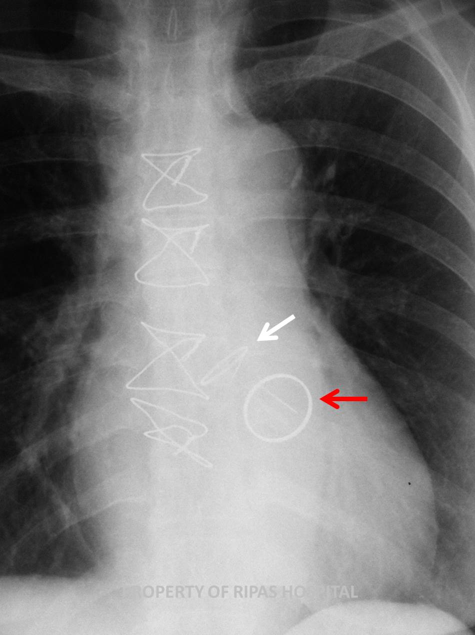
Figure 1: Chest xray of a woman who had undergone
open heart surgery (presence of central sternal wires) with 2 metalic
rings structures inside the heart, one verticle ring and another a
horizontal disc, just above the ring to indicate both mitral and aortic
valves replacement respectively.
(Click on image to
enlarge) |
|
|
|
|
Mitral
stenosis is a condition characterised by a narrowing of the opening of the
mitral valve, resulting in an obstruction of left ventricular inflow tract.
Unlike the aortic valve, the mitral valve consists of 5 structural components
(Annulus, chordae, papillary muscles, left ventricular wall and valvular leaflets) which can be individually
affected causing stenosis but in most cases, are a combination of abnormalities
of 2 or more structures. The aetiologies of mitral valve stenosis can be classified into
2 groups as shown in the table 1 below:
Table
1: Aetiologies of aortic stenosis
|
Neonate/Infant/Children |
Acquired |
|
Congenital mitral stenosis |
Rheumatic mitral stenosis |
|
Cor Triatriatum |
Infective endocarditis with
large vegetation |
|
|
non-rheumatic mitral
annular calcification |
|
|
Rheumatic heart disease |
|
|
Ball valve thrombus |
|
|
malignnant carcinoid disease |
|
|
SLE |
|
|
Rheumatoid arthritis |
|
|
mucopolysaccharidosis of the Hunter-Hurler phenotype |
|
|
Fabry disease |
|
|
Whipple disease |
|
|
Methysergide therapy |
The
natural history of mitral valve stenosis arising from Rheumatic fever is one of
life-long progressive narrowing with
a latent period of 20-40 years in which the patient remains asymptomatic. From
the onset of symptoms to development of disability may take up to 10 years.
Normal mtral valve area is 4.0-5.0 cm2. Symptomatic mitral stenosis
occur when orifice area decreased down to 1.4-2.5 cm2, with critical
mitral stenosis occurring when orifice area is less than 1.0 cm2.
Signs and symptoms of mitral valve stenosis is shown in table 2.
Table
2: Signs and Symptoms of Mitral stenosis
|
Symptoms |
Signs |
|
Dyspnoea |
Loud first heart sound |
|
Fatigue |
Diastolic
murmur |
|
Palpitation |
Opening snap murmur best heard over the apex, which
occurs when the leaflets are mobile. This disappears when the leaflets
are rigid and calcified |
|
Haemoptysis |
Signs of right ventricular failure - RV heave, tricuspid
regurgitation, hepatomegaly, ascites, |
|
Paroxysmal Nocturnal dyspnoea |
Atrial Fibrillation |
|
|
thromboembolic events (20%) |
Mitral
Valve Replacement
Indications for surgery is dependent on the severity of the mitral stenosis.
Asymptomatic patients are not recommended for operation and should be follow up
regularly to monitor progession of their disease. Symptomatic patients who are
otherwise healthy should be advised to undergo surgical correction. Patients
presenting with critical mitral stenosis should undergo urgent operation.
Preoperative preparations
·
Blood
investigations: FBC, Renal panel, LFTs, coagulation screen, cross match.
·
ECG,
Echocardiography to assess severity of stenosis and LV function, TOE for better
assessment particularly of leaflet structures, annulus, chordae and papillary
muscles.
·
Coronary
angiogram to exclude coronary artery disease if indicated.
·
Dental
check -up to repair or extract decayed tooth and resolve all dental caries.
Surgical procedures for mitral valve stenosis can be
classified into 4 types as shown below in table 3:
Table
3: Choice of surgical procedures for mitral valve stenosis
|
Types of procedures |
|
|
1) Catheter based mitral valvotomy |
|
|
2) Open or closed surgical commisurotomy |
|
|
3) Mitral valve repair |
|
|
4) Mitral valve replacement (cases with thick
anterior leaflet, calcification, mitral regurgitation, thick short
chordae) |
mechanical prosthesis, Bioprosthesis (stented
porcine) or mitral homograft. |
As
shown in the chest radiograph Figure 1 and Figure 2 (annotated), this patient
has bivalve replacement using both mechanical St Judes Mechanincal valves as
indicated by the opaque sewing ring and 2 parallel hinge points where the carbon
leaflets are attached.
|
 |
|
Figure 2: Annotated image of Figure 1 with white
arrow pointing at the aortic valve which sits horizontally to the base
of the heart while the mitral valve appears as a ring and sits vertical
to the base of the heart (red arrow).
(Click on image to
enlarge) |
|
|
Survival after MVR
·
Early
(hospital) mortality ranges from 2.5 - 4.0%
·
5-yr
survival – 80%
·
10-yr
survival – 50-80%
Predictors of Survival
·
Coincident coronary artery disease (mortality 6-12%)
·
Left ventricular ejection fraction
Post
operative management
·
Maintain
INR of 2.5 – 3.5 using Warfarin for mechanical valves,
·
For
bioprosthesis, depending on units protocol, may need Warfarin for the first 3
months to allow for the sewing ring to endothelise before stopping Warfarin
·
Cover
with antibiotic Amoxycillin 1.2 g for invasive procedures where breach of
epithelium may occur.
For more information
on:
Mitral stenosis,
please visit this link
http://emedicine.medscape.com/article/155724-overview
Prosthetic heart
valve, please visit this link
http://emedicine.medscape.com/article/780702-overview
Images and text contributed by
Dr Ian Bickle, Department of Radiology,RIPAS Hospital
and
Dr Chong Chee Fui, Department of Surgery, RIPAS
Hospital
All
images are copyrighted and property of RIPAS Hospital.
