IMAGE OF THE WEEK 2014
IMAGE 1
GANGRENE
|
 |
 |
|
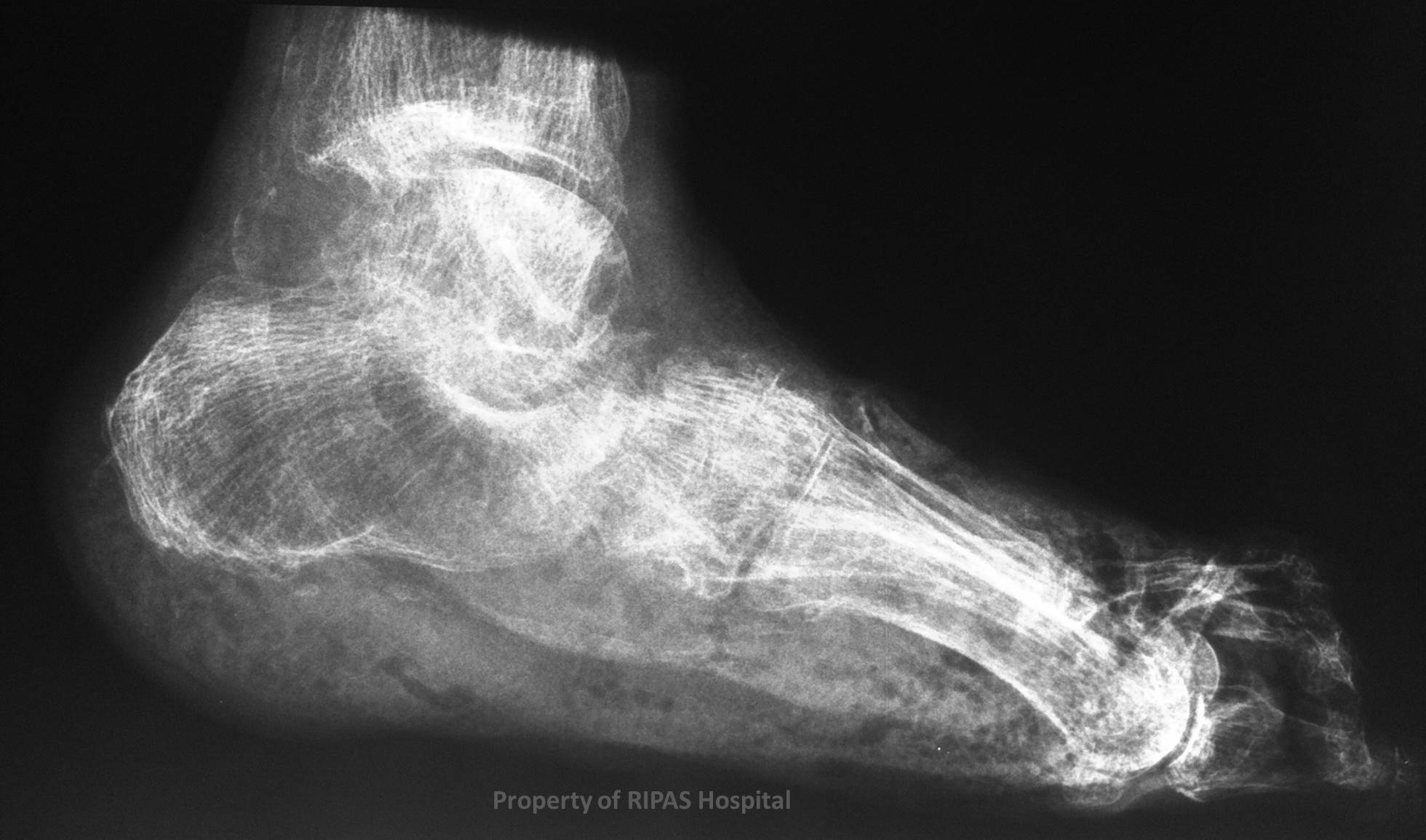
Figure 1: Plain x-ray of the ankle showing multiple
pathologies as annonated in Figure 2.
(Click on image to
enlarge) |
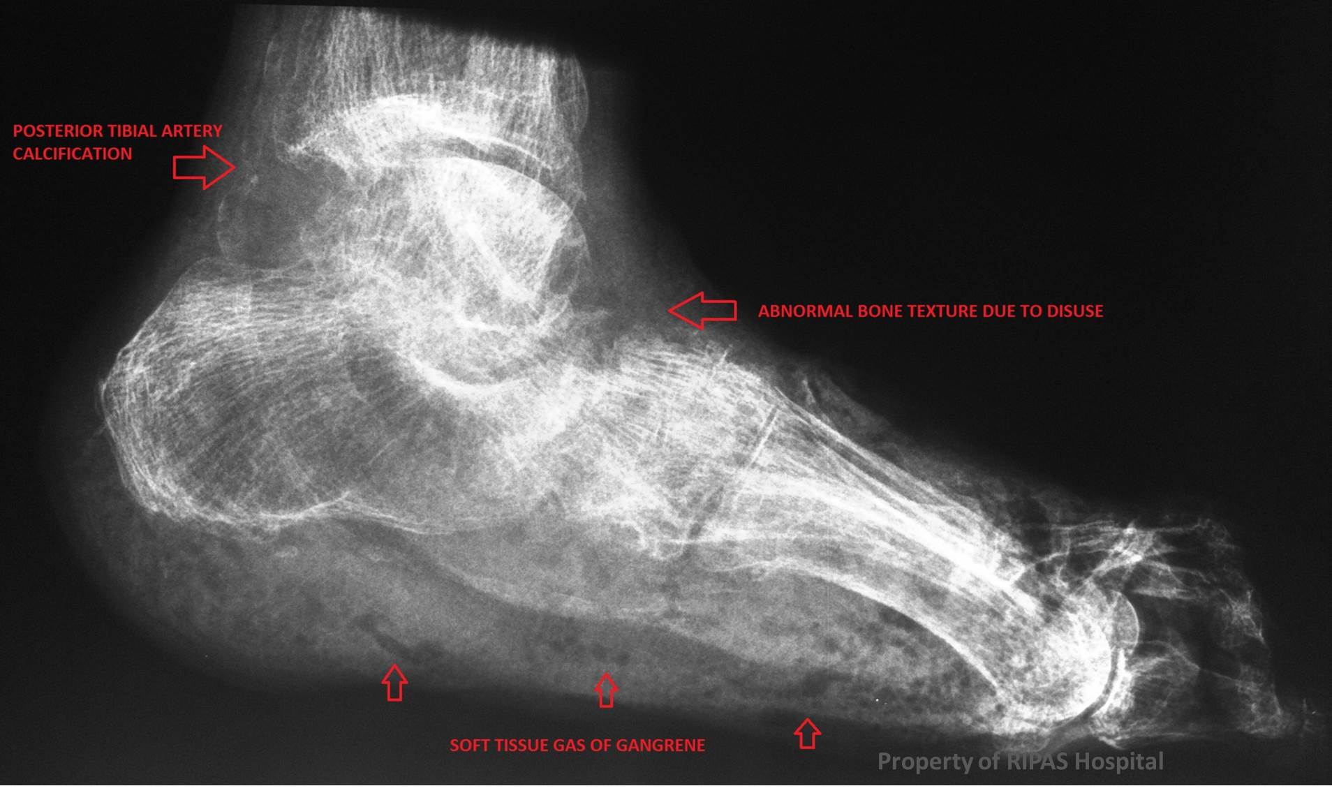
Figure 2: Annotated plain x-ray image of the ankle
of Figure 1 showing disused atrophy, arterial wall calcification and gas
gangrene in soft tissue of the sole.
(Click on image to
enlarge) |
|
|
|
Not everything in medicine necessarily requires an imaging investigation, in
particular a plain x-ray. However, sometimes they are undertaken for an
alternative indication yet reveal findings one must understand and interpret,
despite it perhaps being much more obvious clinically.
In addition, a single x-ray may reveal more than one pathology which are often
related to the same underlying condition.
A classical example of this is a patient with long standing poorly controlled
diabetes, with the multitude of complications which may be set this patient
group.
For example,
-
An ultrasound neck, may reveals a carotid artery stenosis (due to
atherosclerosis) and an enlarged parathyroid gland ( from secondary
hyperparathyroidism.
-
A CT abdomen may demonstrate atrophic kidneys (diabetic microvascular
disease) and the dense bony skeleton of renal osteodystrophy.
-
A plain film of the foot revealing a Charcot’s joint (a result of diabetic
neuropathy ) along with the profound digital artery calcification (due to
atherosclerosis).
-
Or as in this case, a diffusely abnormal texture to the bones of the
foot due to disuse (Sudek’s ) atrophy along with multiple foci of gas in the
soft tissue due to gangrene ( Figure 1 & 2). This is a pre-amputation film
in the extent of disease.
Images and text contributed by
Dr Ian Bickle, Department of Radiology,RIPAS Hospital
All
images are copyrighted and property of RIPAS Hospital.