IMAGE OF THE WEEK 2014
IMAGE 7
EXTRA-AXIAL INTRACRANIAL COLLECTIONS
|
 |
 |
|
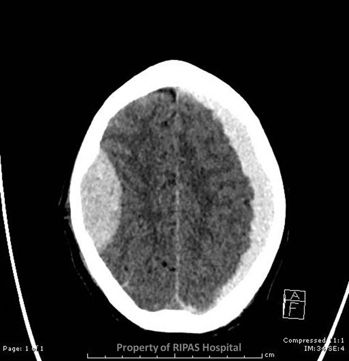
Figure 1: CT head scan showing both an extradural
haematoma on the right and subdural haematoma on the left side. The high
signal intensity indicating an acute bleed.
(Click on image to
enlarge) |
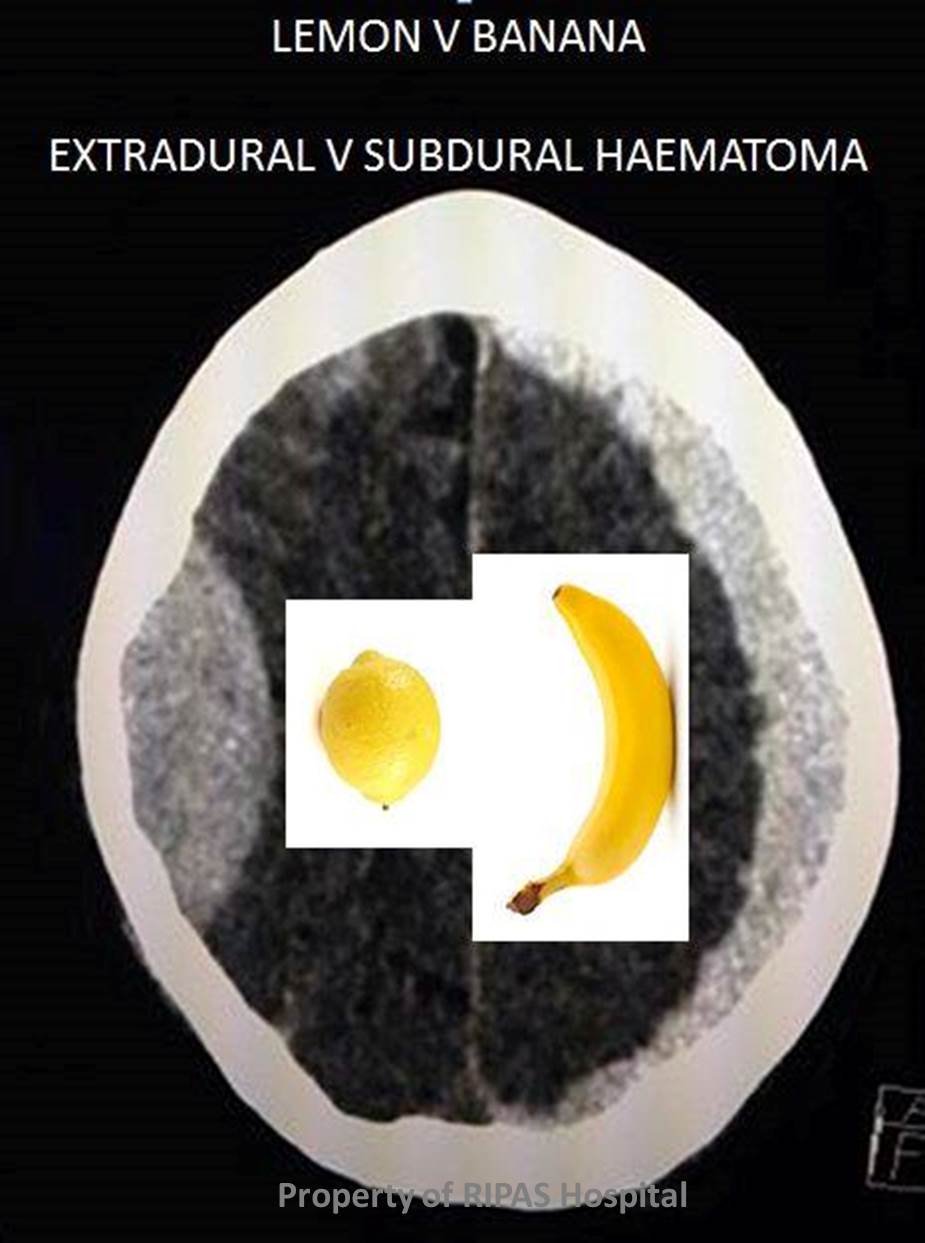
Figure 2: CT head scan from Figure 1a. The
extradural haematoma because of the restriction by the meninges is
shaped like a half lemon as depicted by the picture of the lemon on the
right side while the subdural haematoma without restriction by the
meninges, then to spread out over the surface of the cerebrum and is
shaped like a banana as shown by the banana depicted on the left.
(Click on image to
enlarge) |
|
|
|
One of the most common causes for Out-Of-Hours imaging is head trauma, requiring
the need for a CT head to be undertaken without delay.
Quick, cheap, easy and massively important in management, rapid access CT
imaging is essential in contemporary medicine.
The chief concern of clinicians is BLOOD.
Is there any and does it require neurosurgical input?
Is an operable survivable abnormality present on the CT?
If blood is present, where is it and is it isolated or in several sites?
These are:
-
Extra-axial collections: extradural and subdural haematomas
-
Subarachnoid (Figure 1a and b)
-
Intraparenchymal ( haematoma or contusions )
-
Intraventricular
Of these the extra-axial collections are the most immediate and straightforward
to surgically treat through evacuation with a bore hole, especially if there is
significant mass effect on the brain itself.
An extradural
haematoma
is a collection of blood which forms between the inner surface of the skull and
outer layer of
dura. They are typically lentiform in shape and
hence why the appearance is often referred to as looking like a lemon.
A subdural
haemorrhage is
a collection of blood accumulating in the ‘potentia’l space between
the dura and arachnoid
mater of
the meninges. These appear sickle shaped with the
inner wall of the collection essentially parallel to the bone, hence it has been
referred to as banana shaped.
These illustrations beautifully demonstrate in a pictorial fashion the location
of blood in the different types of collections (Courtesy of www.radiopaedia.org.
Click on link for further information -
http://radiopaedia.org/cases/diagram-intracranial-haemorrhage)
Extra-dural haematomas almost always occur in the context of trauma, with 95%
having an associated underlying fracture. Sub-durals can occur in traumatic and
non-traumatic circumstances, with fractures uncommonly associated.
CT can also assist in aging the extra-axial collection, especially in the case
of subdural haematomas into being: hyperacute, acute, subacute or chronic
depending on the attenuation value and its density in relation to the adjacent
brain parenchyma.
Images and text contributed by
Dr Ian Bickle, Department of Radiology,RIPAS Hospital.
All
images are copyrighted and property of RIPAS Hospital.
