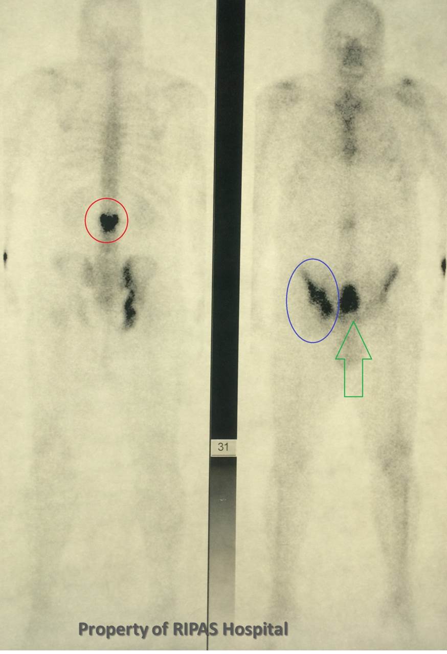
Figure 1: Technetium-99m radioisotope bone scan showing increased uptake in the lumbar spine (hot spot) and delay uptake in the iliac crest and bladder.
(Click on image to enlarge)
IMAGE OF THE WEEK 2014
IMAGE 12
PAGET'S DISEASE
|
|
|
Figure 1: Technetium-99m radioisotope bone scan showing increased uptake in the lumbar spine (hot spot) and delay uptake in the iliac crest and bladder. (Click on image to enlarge) |
Paget’s disease (of the bone) is a chronic disease of the bone, largely inflicting those over the age of 50 years. It results in excessive bony remodelling and may be both asymptomatic and highly problematic depending on its extent and location.
It can occur in virtually any part of the human skeleton; however it is most commonly encountered in the pelvis, spine, long bones and skull.
It is reported to have 3 phases: lytic, mixed and the most frequently sclerotic types. The latter is most commonly identified on imaging investigations.
It may be mono or polyostotic in nature.
The appearances on plain radiograph of the pelvis include:
- Thickening of the bony trabeculae, in particular in the early stages the iliopectineal line
- Sclerotic bony expansion
- Protrusio acetabulae
- Pathological fracture
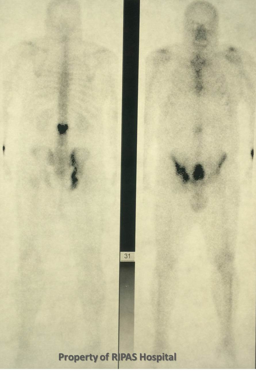
A radioisotope bone scan may visualise the extent of the disease (whether it is polyostotic) and is very sensitive, but not specific. Involved bone will have high tracer uptake on the bone scan, such as the vertebrae and hemipelvis (Figure 1,2 & 3). The main differential is bony metastatic disease.
|
|
|
|
Figure 2: Annotated image of Figure 1 Technetium-99m radioisotope bone scan. (Click on image to enlarge) |
Figure 3: Enlarge image of Figure 2 showing increased uptake in the iliac crest and bladder. (Click on image to enlarge) |
|
|
|
|
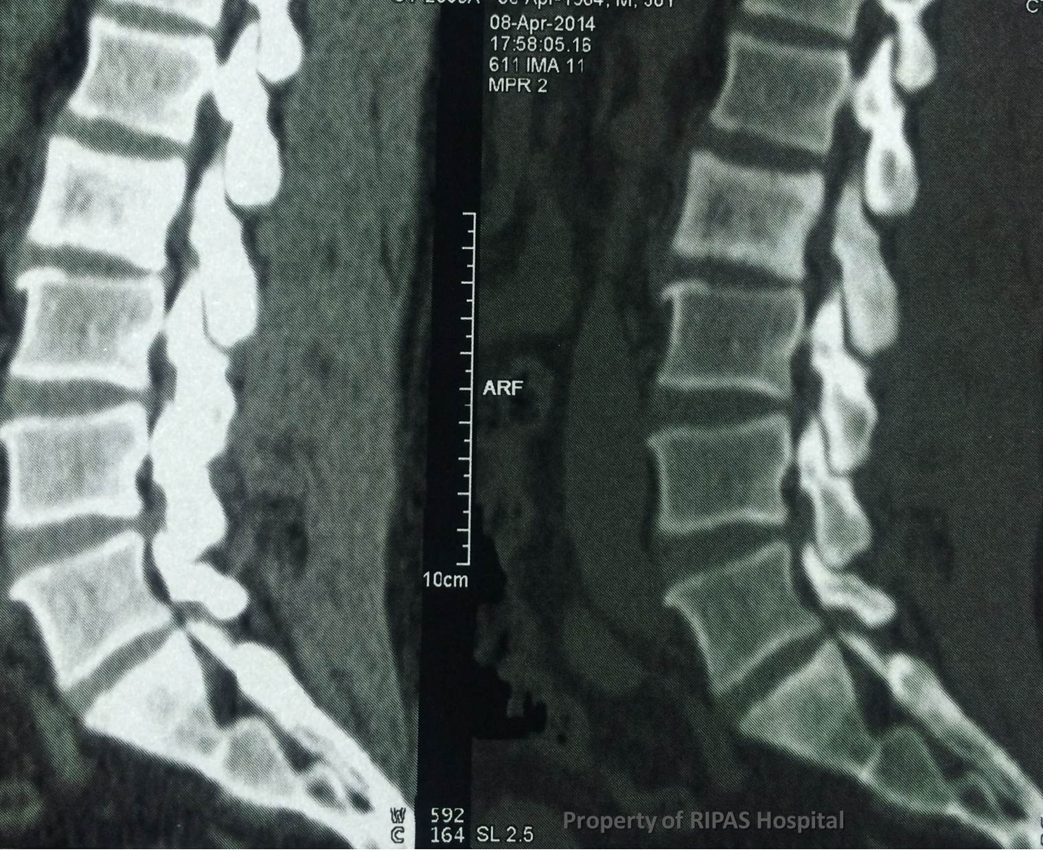
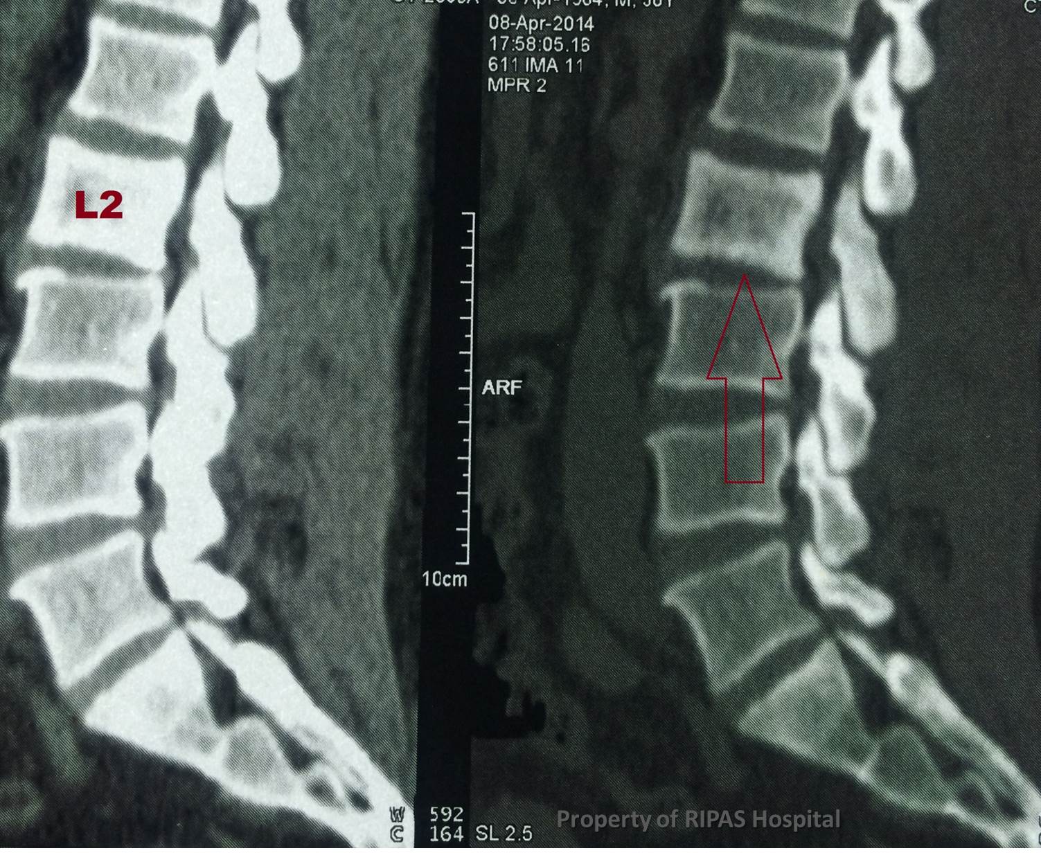
Figure 4: MRI image showing increased density of the L2 lumbar spine body. (Click on image to enlarge) |
Figure 5: Annotated image showing increased density of L2 Lumbar spine body. (Click on image to enlarge) |
Similar appearances can occur in the spine. A single vertebral body may be involved, which is expanded and sclerotic to which the term an ivory vertebra is given (Figures 4 & 5). Metastases and lymphoma can give a similar appearance.
Images and text contributed by
Dr Ian Bickle, Department of Radiology,RIPAS Hospital.
All images are copyrighted and property of RIPAS Hospital.
![]()