IMAGE OF THE WEEK 2014
IMAGE 6
AVASCULAR NECROSIS OF THE FEMORAL HEAD
|
 |
 |
|
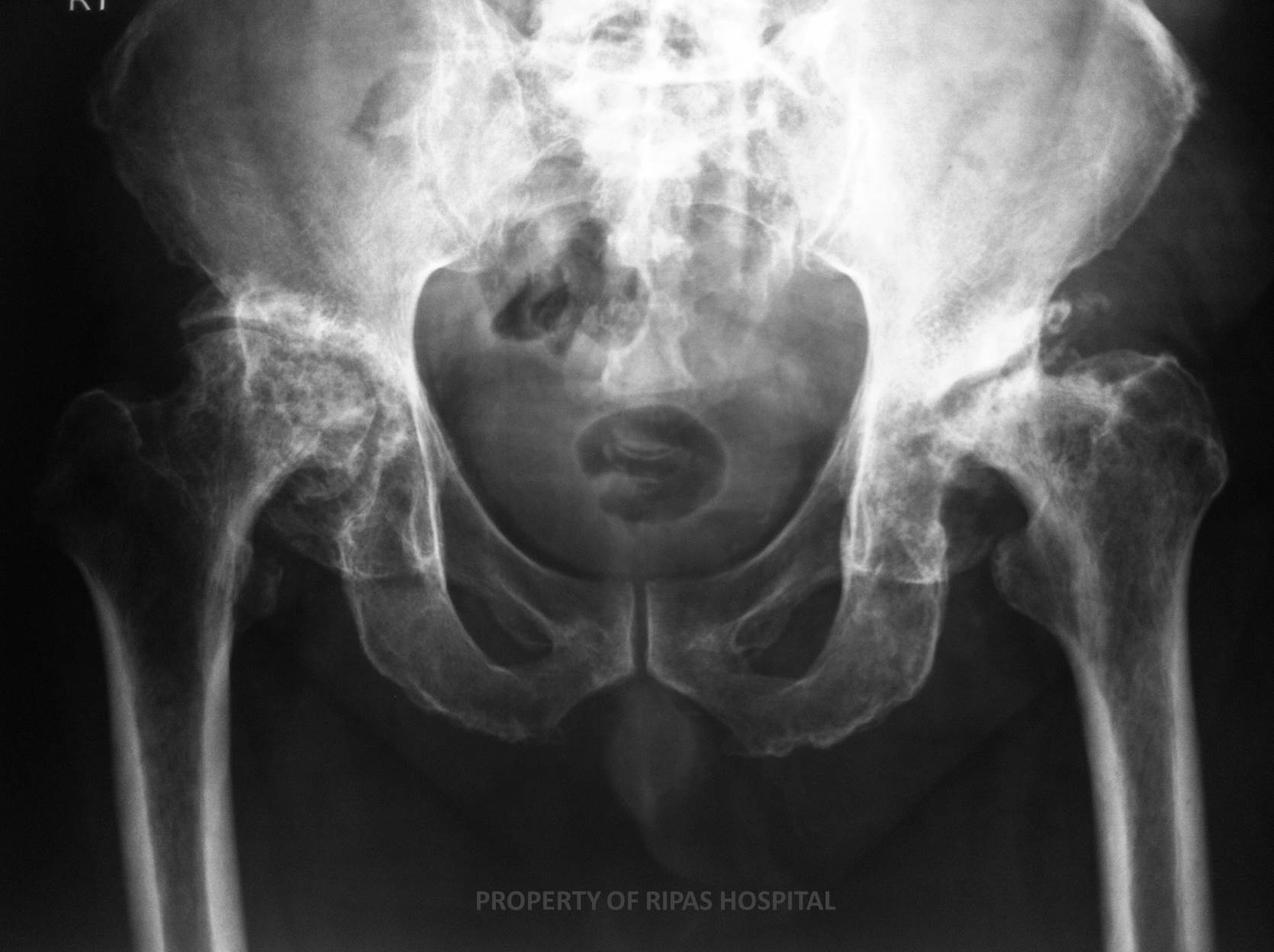
Figure 1: Pelvic xray showing complete destruction
of both femoral heads with loss of articular surface between femoral
head and acetabulum. Both femoral heads appeared irregular and deformed.
(Click on image to
enlarge) |
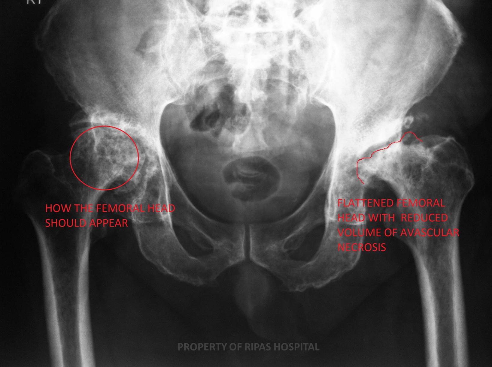
Figure 2: Annotated pelvic xray of Figure 1, showing
complete destruction of both femoral heads with loss of articular
surface between femoral head and acetabulum. Both femoral heads appeared
irregular and deformed. The red ring indicates how the normal femoral
head should appeared in relation to the acetabulum
(Click on image to
enlarge) |
|
|
|
Avascular
necrosis (AVN)
is general term used to describe osteonecrosis resulting from an ischaemic
injury to subchondral bone. A wide range of causes may be responsible for the
ischaemic event arising from a disrupted blood supply. Some causes are more
common, which are indicated by the mnemonic
STARS.
S - steriods
T - trauma
A - alcohol
excess
R – radiation
induced osteonecrosis
S - sickle
cell disease
Although it can potentially occur in any bone, some sites are more prone to AVN,
including the scaphoid, navicular, 2nd metatarsal and femoral head.
Many of these have specific eponymous names, such as Legg-Calve-Perthes disease
in the case of the femoral head.
In the early stages of the disease plain radiographs may be normal, with MRI
being the imaging investigation of choice, with a sensitivity close to 100%.
The double line sign (on T2 weighted sequences) is well documented, which is
pathgnomonic for AVN.
In the later stages of the disease the bone loses volume, collapses and becomes
remodelled, especially in the case of the femoral head (Figure 1 and 2). The
bone appears sclerotic and secondary degenerative changes invariable follow.
Images and text contributed by
Dr Ian Bickle, Department of Radiology,RIPAS Hospital.
All
images are copyrighted and property of RIPAS Hospital.
