IMAGE OF THE WEEK 2014
IMAGE 4
PATHOLOGICAL FRACTURES
|
 |
 |
|
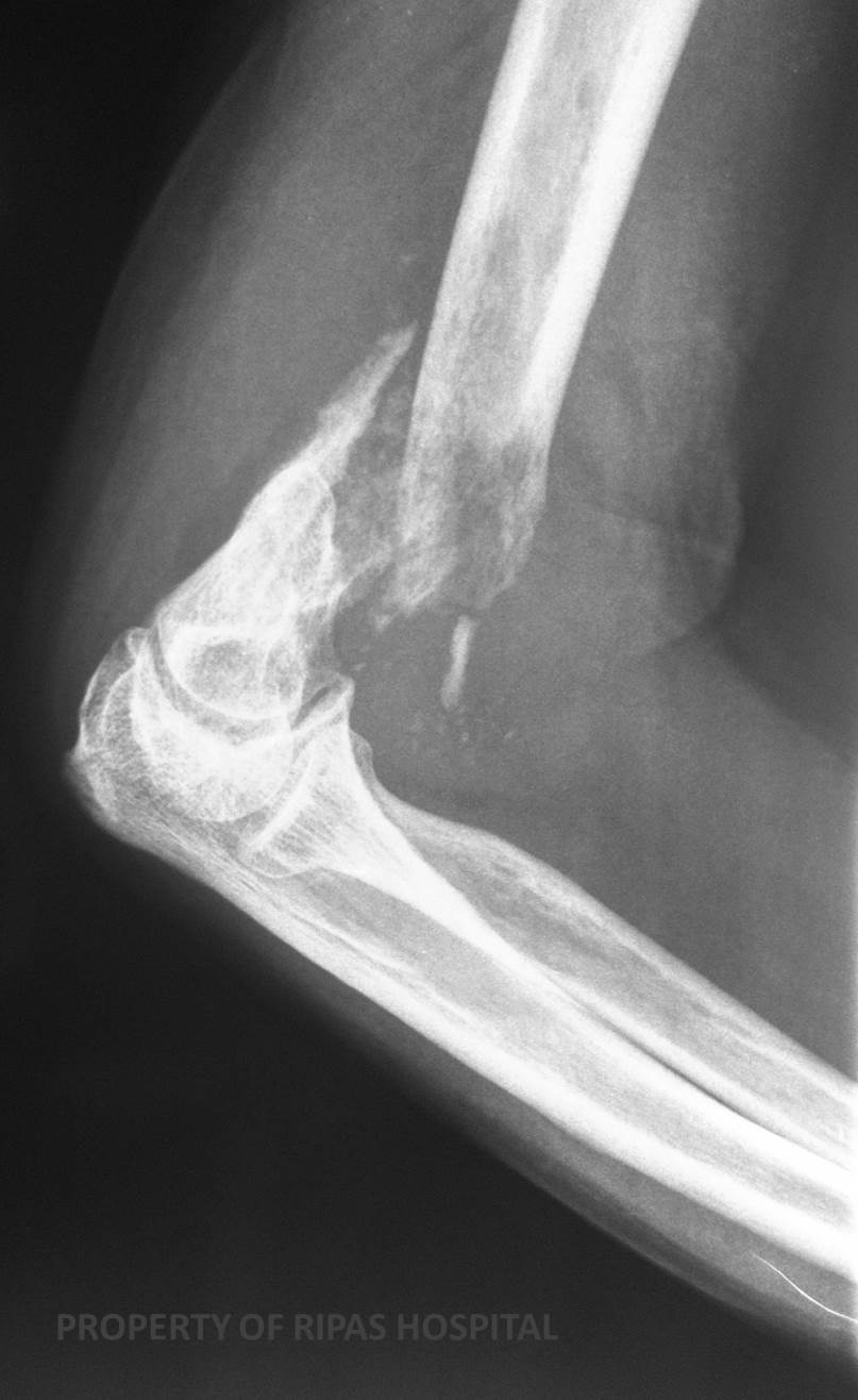
Figure 1: Lateral xray of the elbow showing a
pathological fracture through the distal part of the humerus bone. The
fracture site is not clean but had multiple fragments with edges of the
bone appearing radiolucent due to metastasis present in the bone.
(Click on image to
enlarge) |
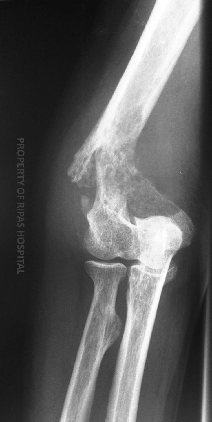
Figure 2: AP view of the smae xray of the elbow,
showing the same fracture with anterior and lateral displacement of the
upper part of the humeral bone.
(Click on image to
enlarge) |
|
|
|
PATHOLOGICAL FRACTURE
The majority of fractures
are simple and the result of trauma. A small percentages (< 1%) are
pathological fractures.
Pathological fractures commonly
occur in abnormal bone. The underlying abnormality may be malignant or
non-malignant in nature.
The term is usually assigned to fractures than occur through a focal bone
abnormality. In some cases, the initial presentation of an unknown malignancy
is following a low impact fracture, which is subsequently identified on
imaging. A focal bone lesion may be identified or a generalised abnormality of
the bone texture at the site of the fracture.
Causes
Malignant
Metastasis: The commonest primaries to metastasize to bone are: lung, breast,
renal.
Multiple myeloma
Other primary bone malignancies, such as osteosarcoma
Non Malignant
Generalised
Osteoporosis – the commonest
Osteogenesis Imperfecta
Osteomalacia
Renal
Osteodystrophy
Gaucher's Disease
Localised
Osteomyelitis
Pagets
Disease
Fibrous Dysplasia
Fibrous Histiocytoma
Giant
Cell Tumour of Bone
Non-ossifying fibroma
In this case, a CT chest,
abdomen and pelvis was performed, which identified a 1.2cm spiculated lung
mass. This was not technically amenable to CT guided biopsy. The final
histopathological diagnosis was a squamous cell carcinoma of the lung,
ascertained from tissue at the fracture site at the time of open reduction and
internal fixation (ORIF).
|
 |
|
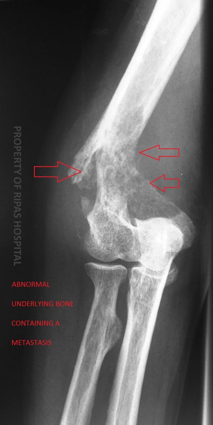
Figure 3: Annotated xray of the elbow in Figure 2,
showing the same fracture with anterior and lateral displacement of the
upper part of the humeral bone.
(Click on image to
enlarge) |
Images and text contributed by
Dr Ian Bickle, Department of Radiology,RIPAS Hospital
