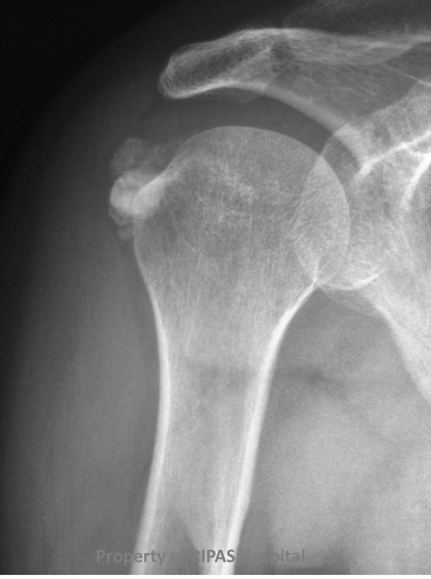
Figure 1: Calcification of the tendons of the shoulder (right) -Calcifictendinitis.
(Click on image to enlarge)
IMAGE OF THE WEEK 2014
IMAGE 14
CALCIFICTENDINITIS
|
|
|
|
Figure 1: Calcification of the tendons of the shoulder (right) -Calcifictendinitis. (Click on image to enlarge) |
|
The rotator cuff of the shoulder is composed of 4 tendons which are responsible for different movements of the shoulder.
Supraspinatus – abduction
Infraspinatus - external rotation
Teres minor - external rotation
Subscapularis - internal rotation
These tendons are prone to inflammation, which is a self limiting, but problematic, condition due to deposition of calcium hydroxyapatite crystals within tendons the rotator cuff, typically the supraspinatus (Figure 1).
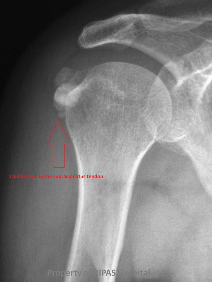
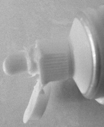
The appearances of ‘blobs’ of calcification in the supraspinatus tendon has been described as appearing like toothpaste squeezed from a tube (Figure 2 & 3).
|
|
|
|
Figure 2a: Magnified image of figure 1 showing the tendon calcifications at the right shoulder joint. (Click on image to enlarge) |
Figure 2b: Annotated image of Figure 2a with red arrow pointing at the calcification of the tendons at the right shoulder joint. (Click on image to enlarge) |
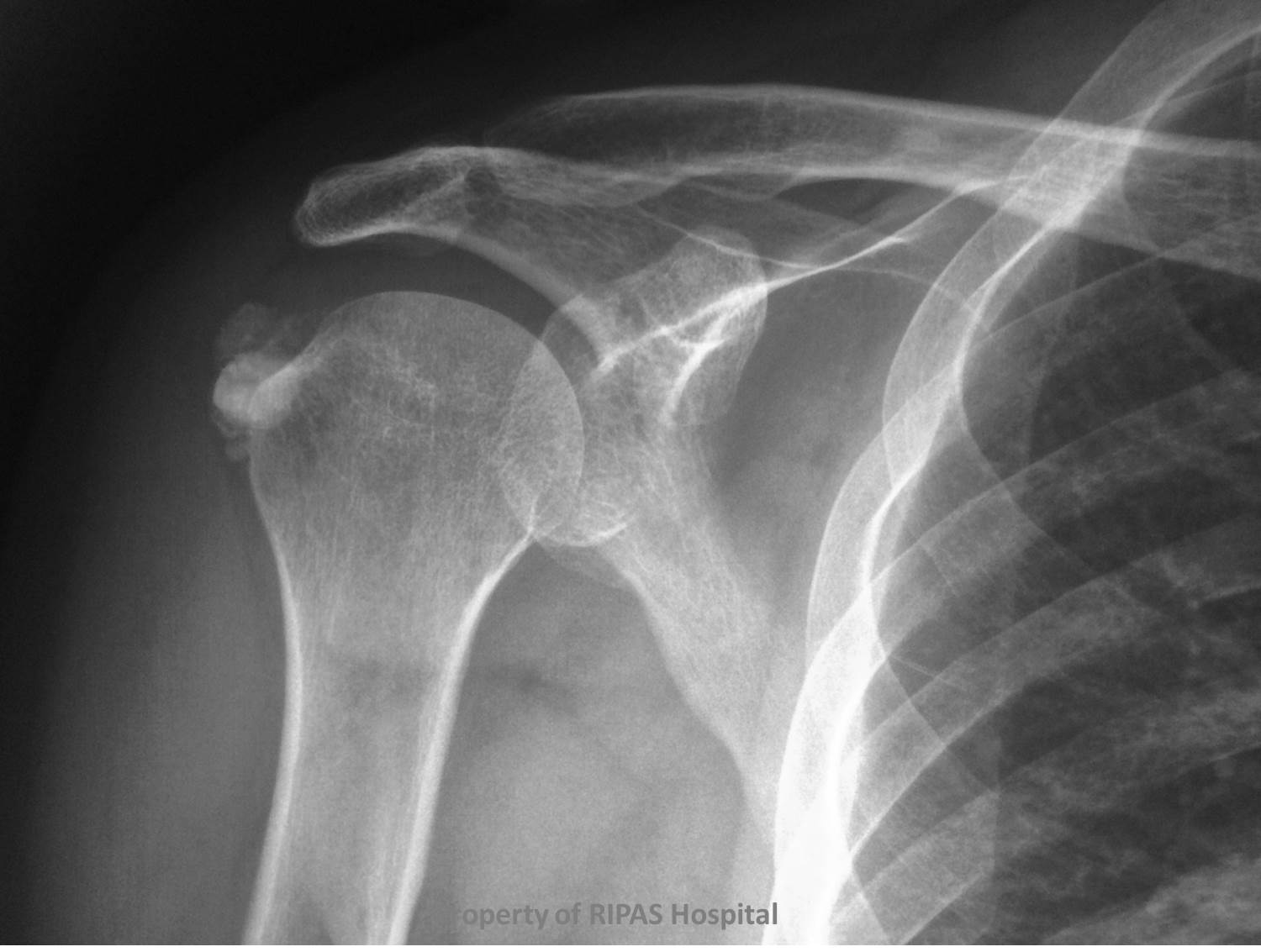
This can be identified on plain radiograph, ultrasound, MRI and CT of the shoulder.
Rotator cuff pathology is typically assessed with plain radiograph and ultrasound first-line with MRI reserved for more problematic cases.
|
|
|
Figure 3: Calcifictendinitis of the tendons of the shoulder joint as depicted above resembles a blob of toothpaste being squeezed out of the tube. (Click on image to enlarge) |
Images and text contributed by
Dr Ian Bickle, Department of Radiology,RIPAS Hospital.
All images are copyrighted and property of RIPAS Hospital.
![]()