Figure 1: Normal xray of the foot
(Click on image to enlarge)
IMAGE OF THE WEEK 2014
IMAGE 8
OSTEOMYELITIS OF THE FOOT - TIME LAPSE XRAY CHANGES
|
|
|
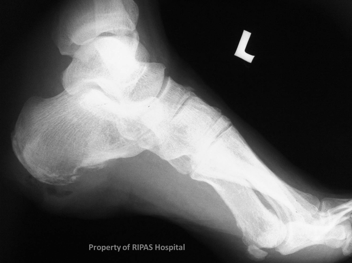
Figure 1: Normal xray of the foot (Click on image to enlarge) |
Osteomyelitis is infection of bone - it can occur in any bone and the imaging appearances are identical.
Certain patient groups are particularly susceptible to osteomyelitis - the single biggest group being those with long standing diabetes mellitus. This is a particular problem in the feet, given the combined problem of:
a. Susceptibility to infection
b. Poor circulation - largely through arterial atherosclerosis
c. Peripheral neuropathy
The lack of sensation arises from diabetic peripheral neuropathy allows repeated trauma and an absence of pain, contributing further to the bony destruction through late presentation to medical attention.
Imaging usually begins with plain radiographs, despite their relative lack of sensitivity. MRI is the most sensitive modality allowing bone oedema to be illustrated before frank bony destruction has occurred. CT assists in outlining the fine detail of bony destruction.
This case highlights the progressive nature of osteomyelitis and the spectrum of findings on imaging over an 18 month period.
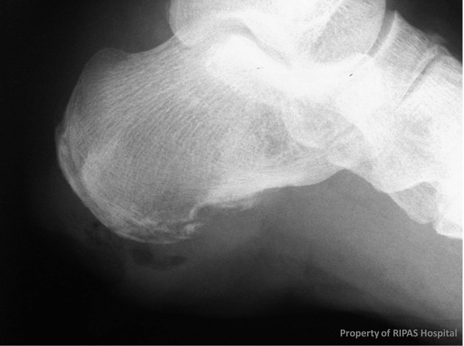
The calcaneum at the start of this patient journey is normal (Figure 1). The bony cortex is intact with no visible abnormality.
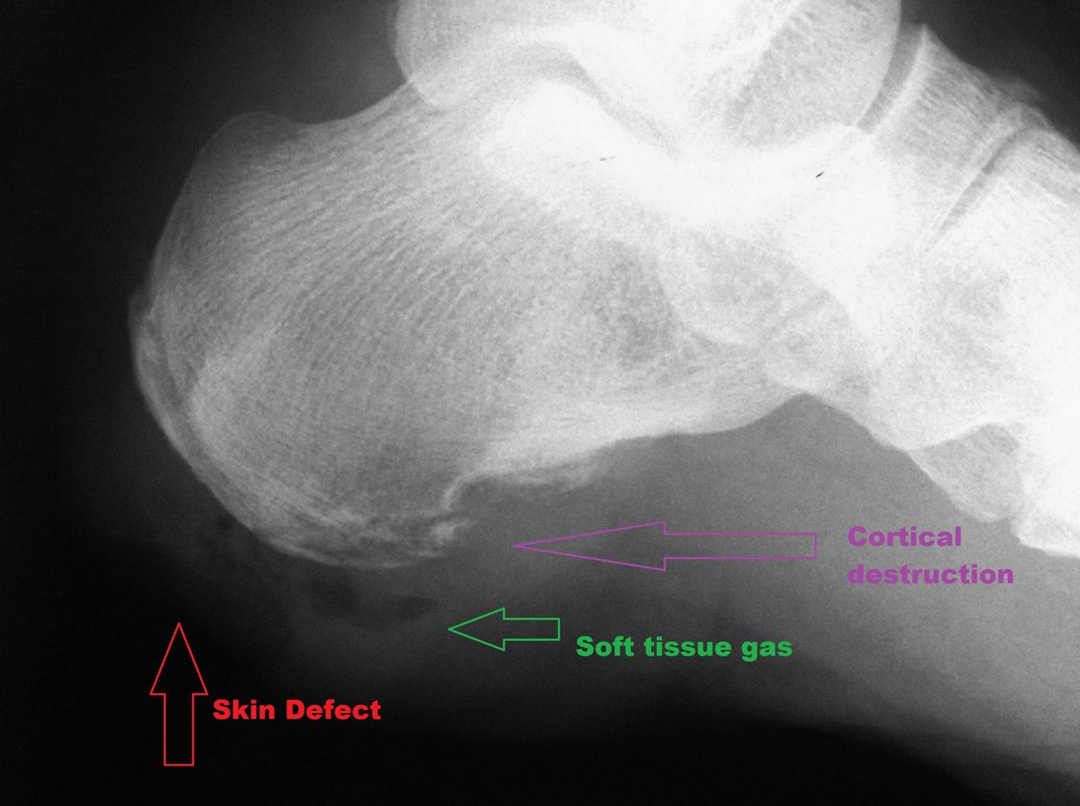
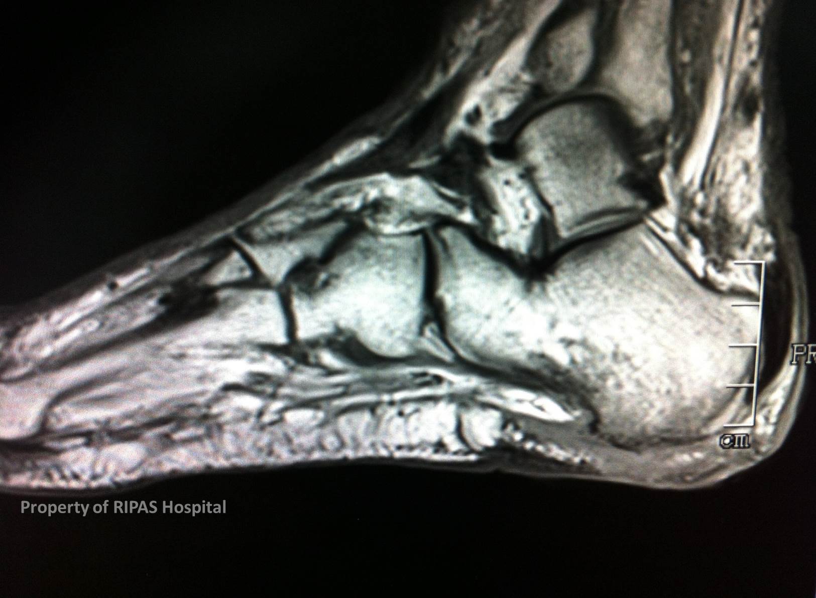
Several months later the skin defect over the heel is apparent, with subcutaneous gas indicative of infection. The cortex of the adjacent calcaneum is now breeched (Figures 2A-C). This is demonstrated in more detail on the MRI (Figure 3), which illustrates the sensitivity of this modality.
 |
|
|
|
Figure 2a (Click on image to enlarge) |
Figure 2b (Click on image to enlarge) |
Figure 2c (Click on image to enlarge) |
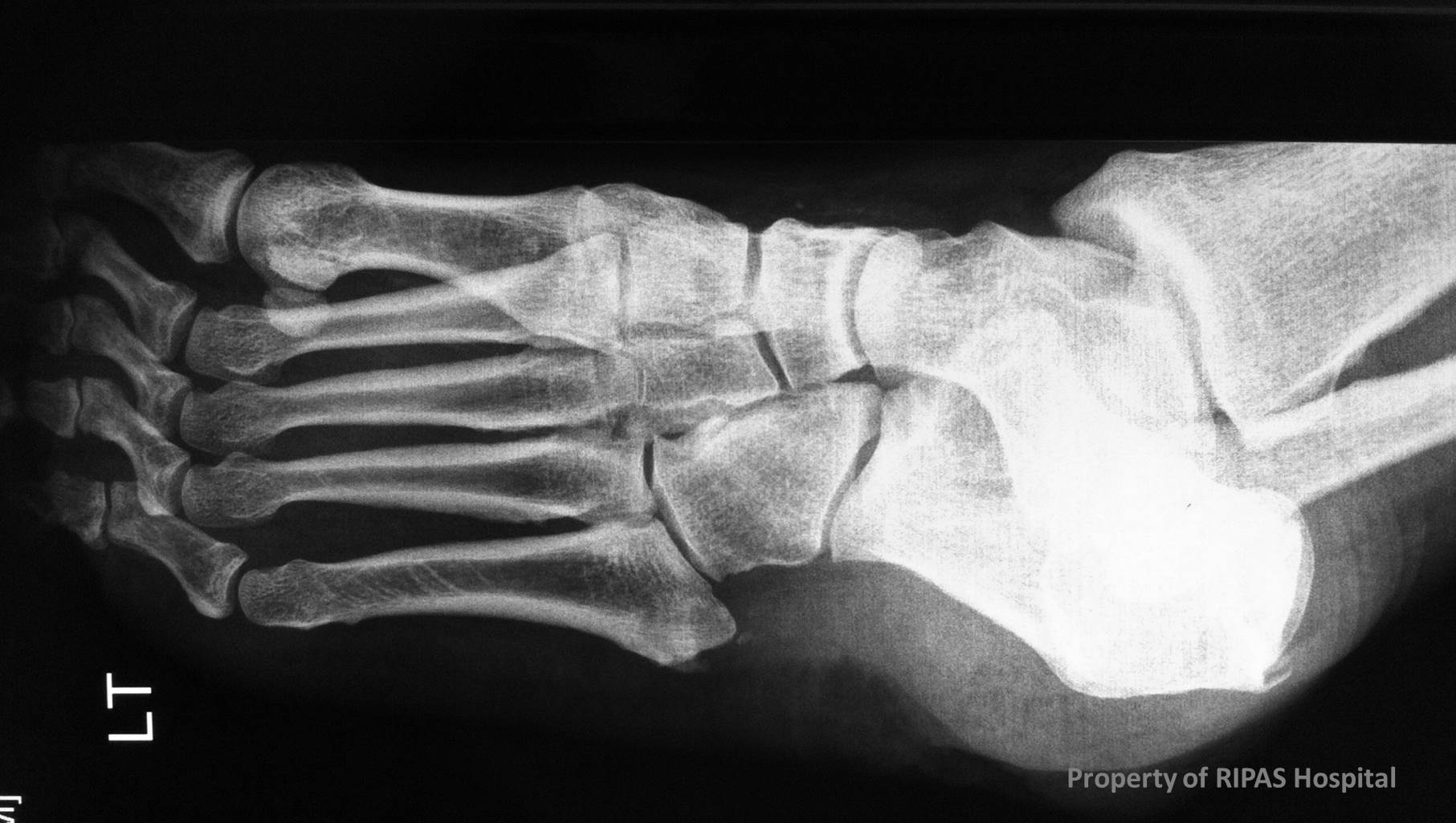
A further period of several months passed. On this occasion the radiographs show a pronounced progression with extensive bony destruction of the cortex and deeper bone marrow (Figure 4).
A further MRI was undertaken soon after, in which the magnitude of destruction is evident, along with infected adjacent periarticular soft tissue and thickening of the Achilles insertion (Figure 5).
|
|
Figure 3 (Click on image to enlarge)
|
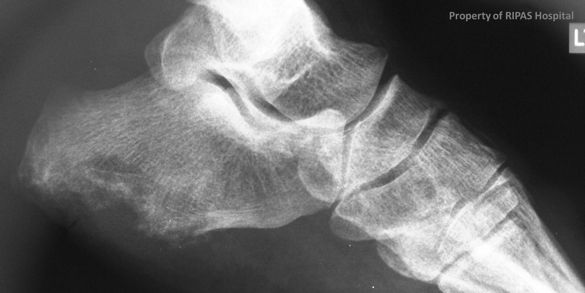 |
Figure 4 (Click on image to enlarge)
|
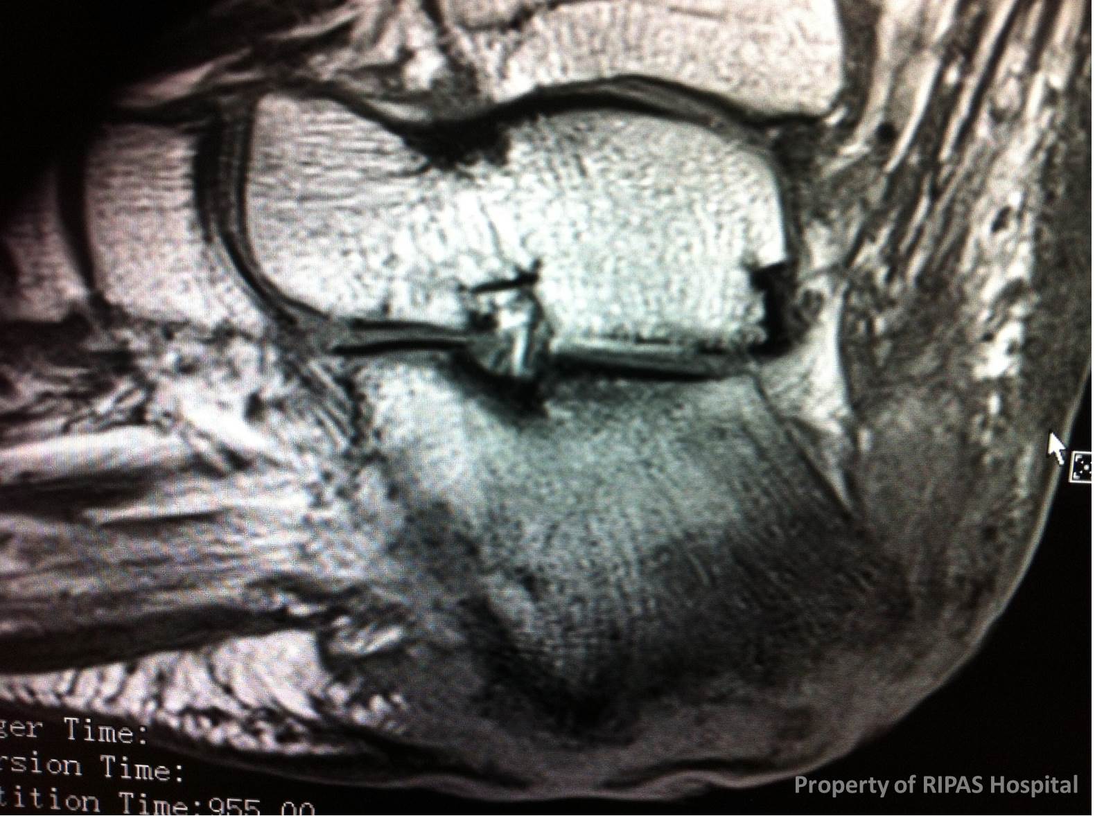 |
Figure 5 (Click on image to enlarge)
|
Images and text contributed by
Dr Ian Bickle, Department of Radiology,RIPAS Hospital.
All images are copyrighted and property of RIPAS Hospital.
![]()