IMAGE OF THE WEEK
WEEK 10
|
|
|
SPINAL BIFIDA
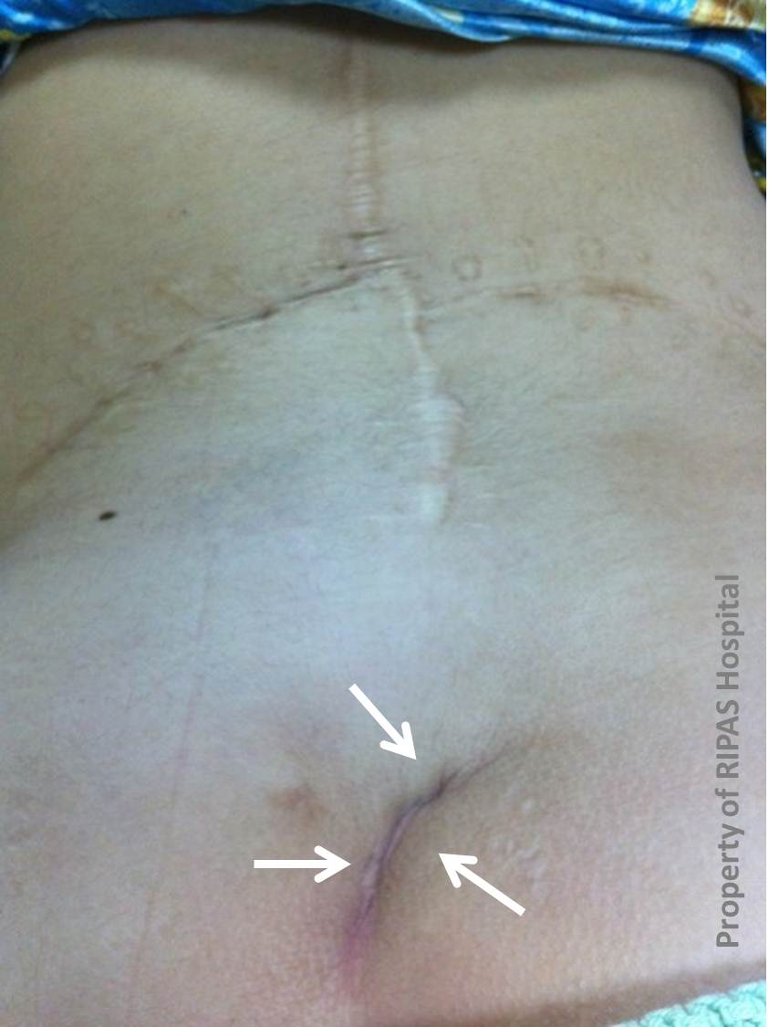
Spina bifida (“cleft spine”) is a birth defect affecting the spinal column. Spina bifida progresses from a cleft, or splitlike opening, in the back part of the backbones (the spinal vertebrae). Spina bifida is the most common of a group of birth defects known as neural tube defects, which affect the central nervous system (brain and spinal cord). An abnormal tuft of hair, a collection of fat, a small dimple (White arrows) or a birthmark on the newborn's skin above the spinal defect may be the only visible clinical sign of the condition.
There are 3 types of spina bifida:
1. Spina bifida occulta: which is hidden, and the defect is not visible. It is rarely linked with complications or symptoms. 2. Meningocele: In this type, the membrane that surrounds the spinal cord may enlarge, creating a lump or “cyst.” This is often invisible through the skin but causes no problems. 3. Spina bifida cystica or also known as myelomeningocele is the most complex and severe form of spina bifida. It usually involves neurological problems which can be very serious or even fatal. A part of the affected spinal cord and nerves arising from the spinal cord are exposed and visible on the outside of the body or they may be enclosed by a cyst.
TREATMENT
Children with spinal bifida occulta seldom require any treatment.
Most children with severe types of spina bifida require a series of operations, which is usually performed within the first 1 to 2 days after birth. At the first surgery, the spine cord is pushed back into the vertebral canal and the defect is closed to prevent infection and protect the spine cord.
Subsequent surgeries are performed to correct the associated deformities (note the scar along the spine of the case above). Urological surgery is often necessary to correct associated renal anomalies (note the left lumbar scar of the case above).
Images prepared by Dr Eddy Ngatemo, Department of Accident & Emergency Medicine, RIPAS Hospital, Brunei Darussalam.
Edited by Mr CF Chong.
All images are copyrighted and property of RIPAS Hospital.
![]()