Figure 1: Click on image to enlarge
Figure 2: Click on image to enlarge
IMAGE OF THE WEEK 2012
WEEK 11
BLADDER RUPTURE
|
|
|
|
|
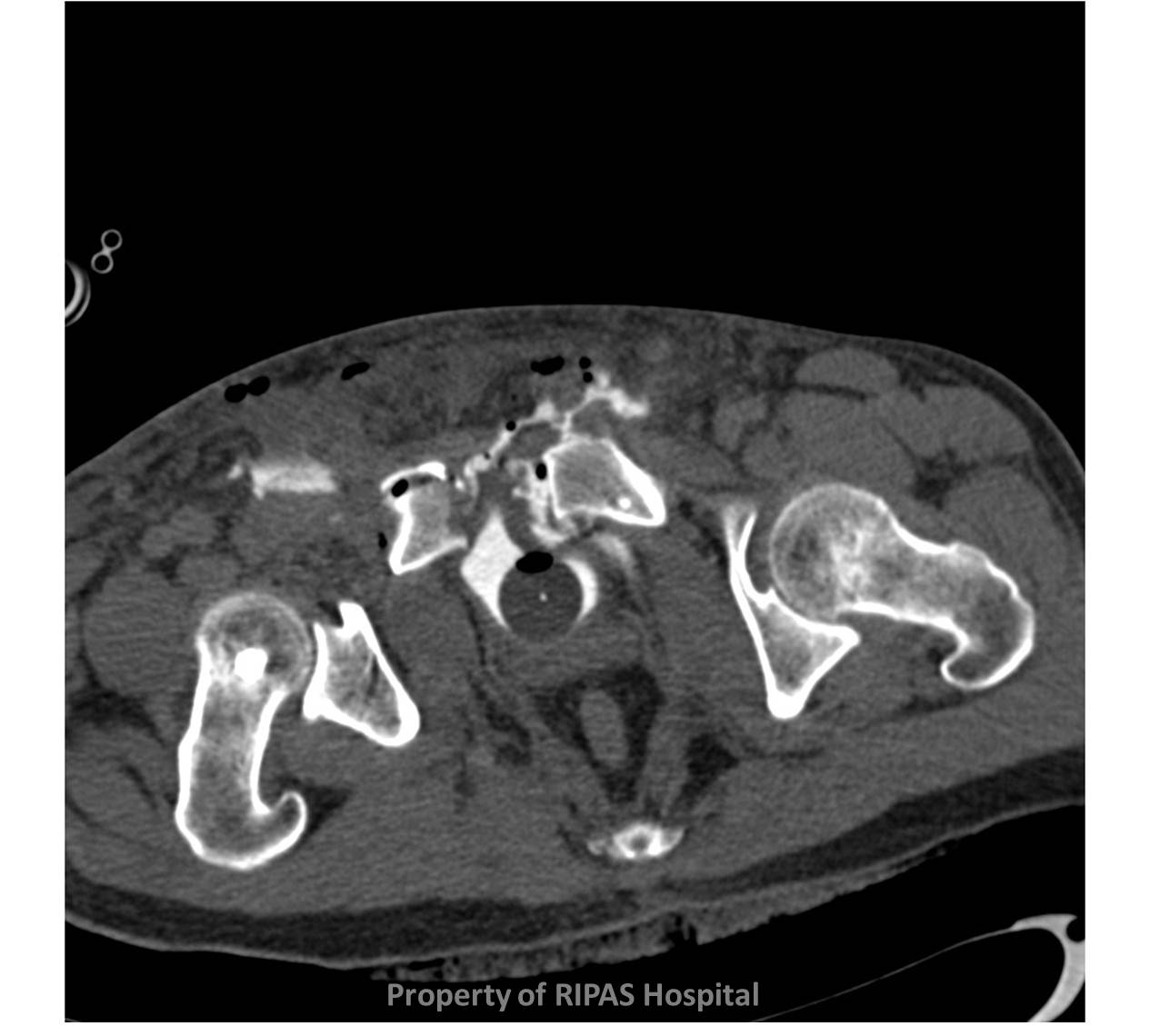
Figure 1: Click on image to enlarge |
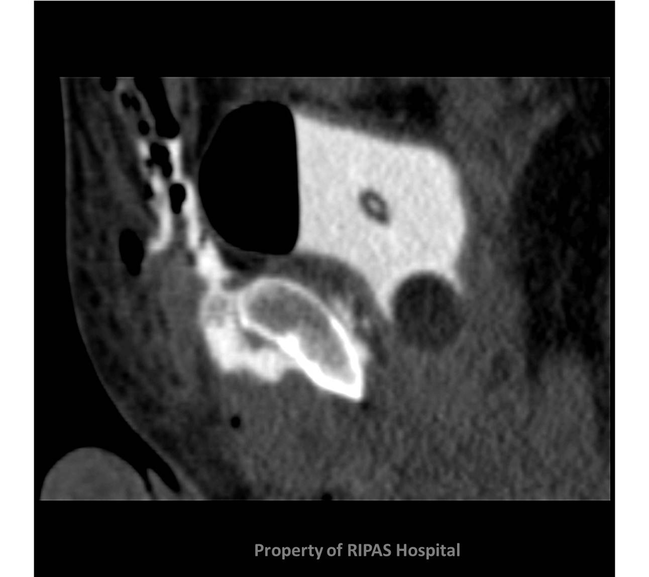
Figure 2: Click on image to enlarge |
|
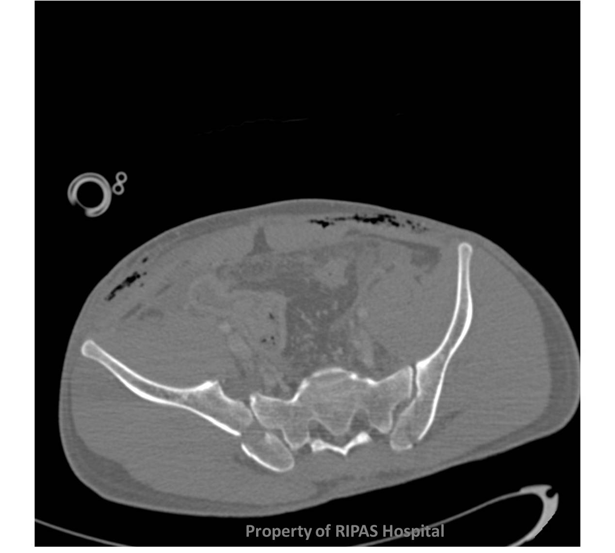
Bladder rupture is uncommon. It can be divided broadly into intra-peritoneal and extra-peritoneal rupture, the latter being more common, representing at least 80% of all cases.
Bladder rupture is commonly associated with severe pelvic fractures, such as those following high speed RTA’s or other high velocity trauma.
Intra-peritoneal rupture
Extra-peritoneal rupture
The diagnosis can be made with either a traditional fluoroscopic cystogram study or with CT cystogram/IVU, the latter being more sensitive.
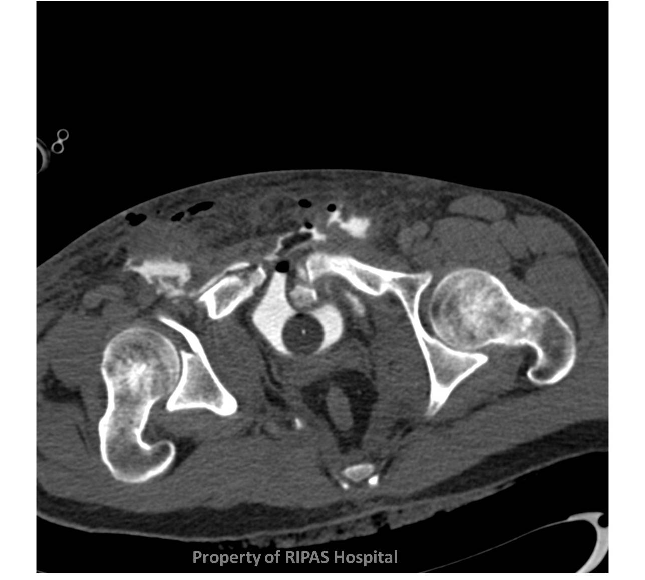
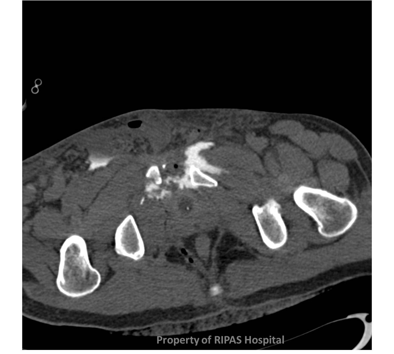
In this case contrast can be seen to extend beyond the confines of the urinary bladder, into the extra-peritoneal space (Figure 1). The leak, as is often the case, is arising from the bladder base, in close proximity to the disrupted pubic symphysis (Figure 2). Further contrast extravastation has occurred into the soft tissues of the anterior abdominal wall/groins bilaterally (Figure 3 & 4). There is an extensive pelvic fracture as is normally the case with extra-peritoneal bladder injuries (Figure 5).
|
|
|
 |
|
Figure 3: Click on image to enlarge |
Figure 4: Click on image to enlarge |
Figure 5: Click on image to enlarge |
Image and text contributed and prepared by
Dr Ian Bickle, Department of Radiology, RIPAS Hospital, Brunei Darussalam.
All images are copyrighted and property of RIPAS Hospital.
![]()