Figure 1: Click on image to enlarge
Figure 2: Click on image to enlarge
Figure 3: Click on image to enlarge
IMAGE OF THE WEEK 2012
WEEK 12
Tuberculous Spondylodiscitis (Pott’s Disease)
|
|
|
|
|
Figure 1: Click on image to enlarge |
Figure 2: Click on image to enlarge |
Figure 3: Click on image to enlarge |
Pott’s disease is named after Sir Percivil Pott, an English surgeon, who was the first to describe tuberculous spondylitis in the late 18th Century. Interestingly it was also Pott who also identified the association between scrotal cancer and soot exposure in chimney sweeps!
Tuberculosis is a true multisystem disorder, and although most commonly associated thought of as a respiratory disease it may affect other body systems, including both the spine and brain. Often this occurs in conjunction with pulmonary disease, but not always. Bone and soft-tissue tuberculosis accounts for approximately 10% of extra-pulmonary tuberculosis cases and between 1% and 2% of total cases.
Radiological Features
Typically involves the lower thoracic vertebrae
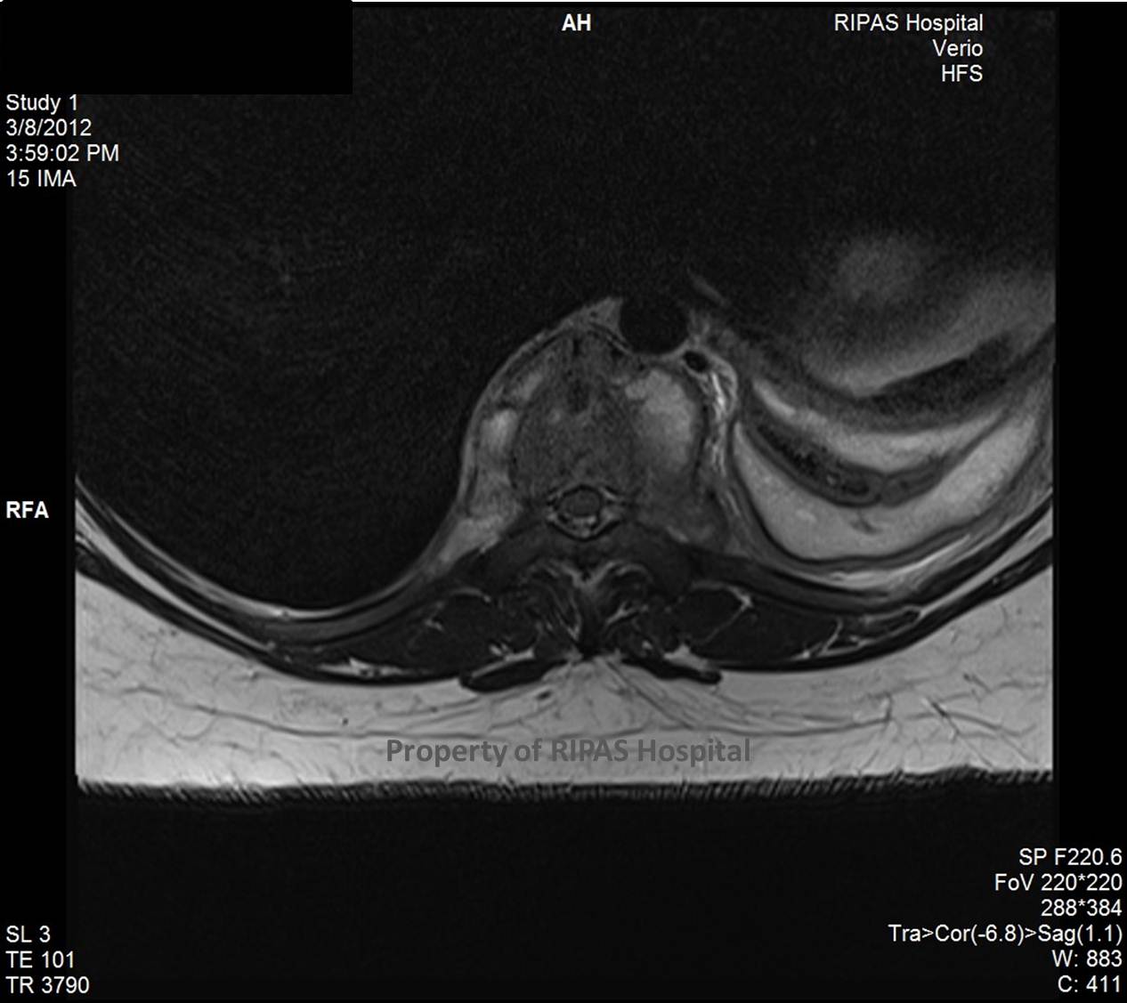
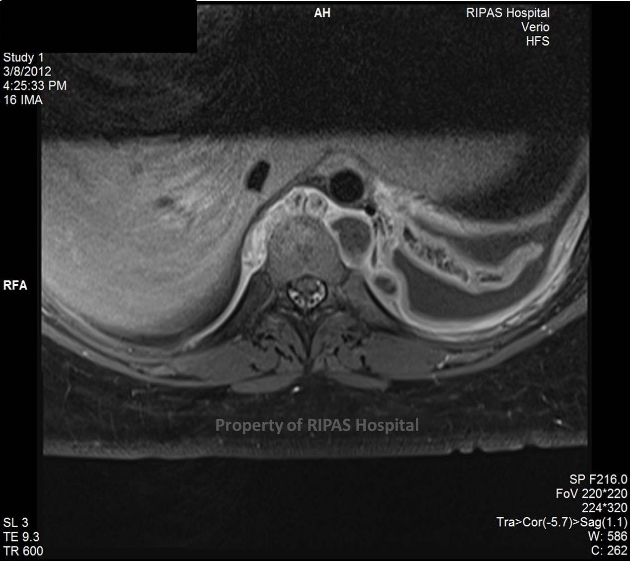
Is commonly associated with a significant paraspinal abscess/inflammatory soft tissue
May cause a gibbus deformity
May be epidural soft tissue and or epidural abscess
May have associated psoas abscess formation
May be multifocal
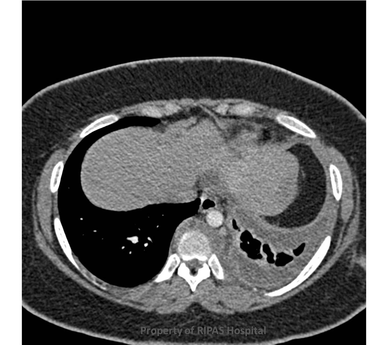
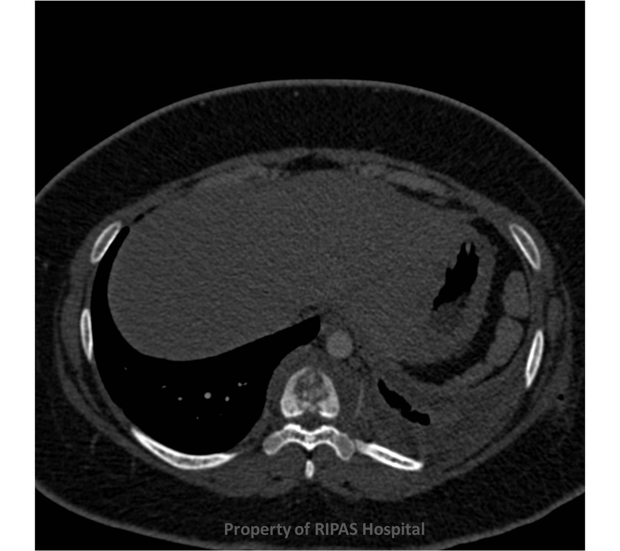
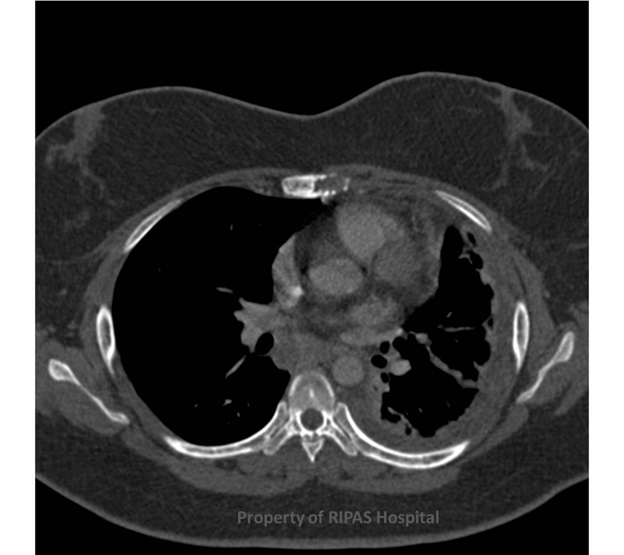
CT may be initially performed and identify bony destruction, such as in this case. There is a T8/9 spondylodiscitis (Figure 1) with associated paraspinal soft tissue (Figure 2), which was associated with destruction of the sternal body (Figure 3).
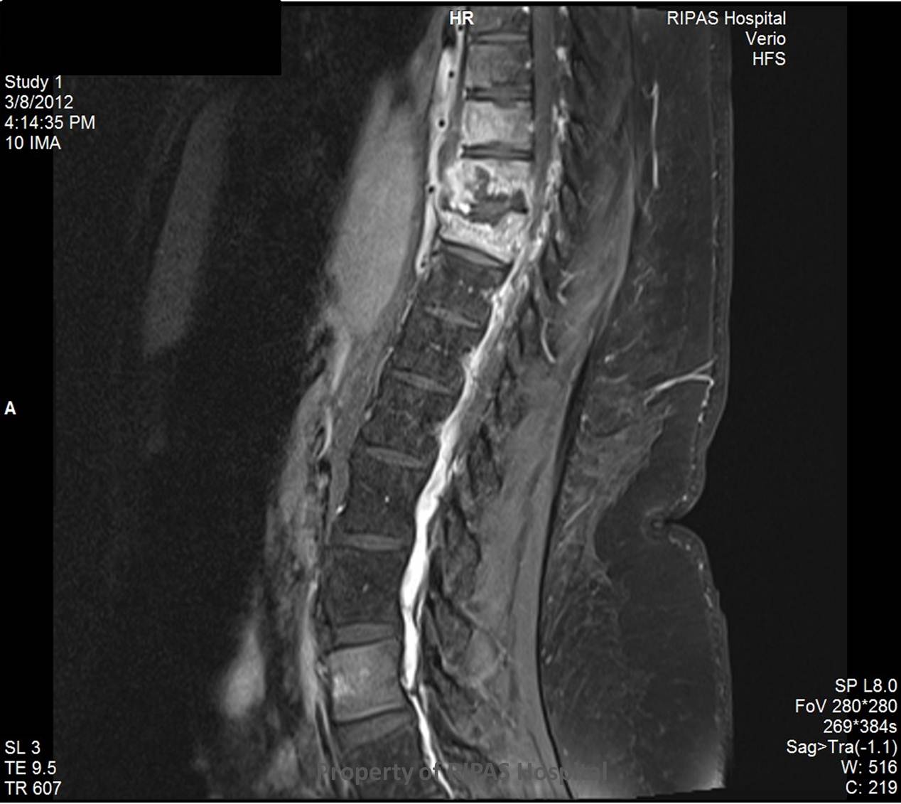
MRI is the investigation of choice. This should include sequences following contrast administration. It is most sensitive at detecting the spondylodiscitis and examining its extent.
The following is seen in this case:
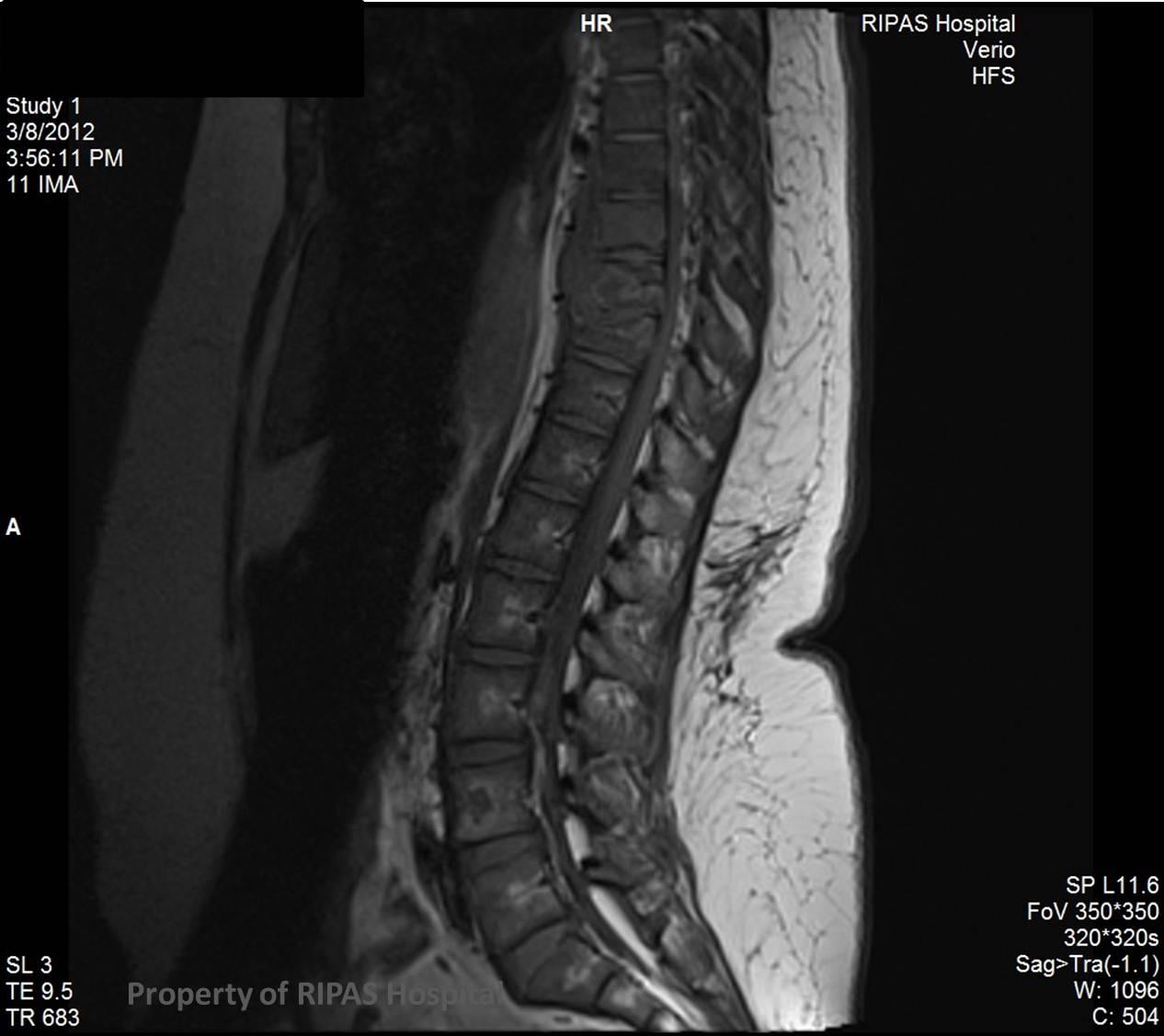
a. Abnormal signal within the bone marrow on T1/TIRM sequences (Figure 4)
b. High signal in the intervertebral disc on the T2 sequences (Figure 5)
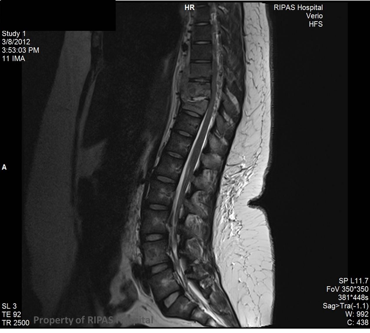
c. Paraspinal soft tissue/collection on T2 (Figure 6)
d. Avid enhancement of the disc/paraspinal tissue on T1 (fat saturation) post contrast administration (Figure 7 & 8).
|
|
|
|
Figure 4: Click on image to enlarge |
Figure 5: Click on image to enlarge |
|
|
 |
|
Figure 6: Click on image to enlarge |
Figure 7: Click on image to enlarge |
|
|
|
|
Figure 8: Click on image to enlarge |
Image and text contributed and prepared by
Dr Ian Bickle, Department of Radiology, RIPAS Hospital, Brunei Darussalam.
All images are copyrighted and property of RIPAS Hospital.
![]()