IMAGE OF THE WEEK
WEEK 13
|
|
|
|
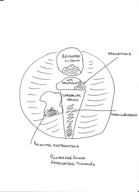
MEDULLOBLASTOMA
Medulloblastoma are included in the histopathological group of the Primitive Neuroectodermal Tumours (PNET) and account for approximately 7-8% of all intracranial tumors and 30% of paediatric brain tumors.
Three quarters of all cases occur in children, with a median age of 9 years, with an incidence of 1.5-2 cases per 100,000 population.
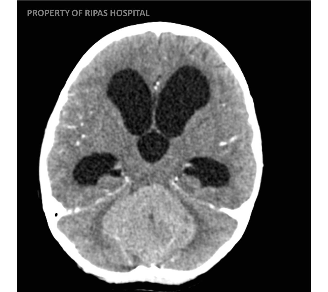
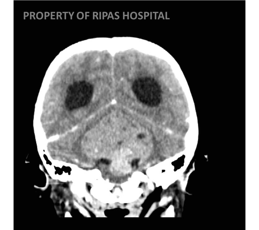
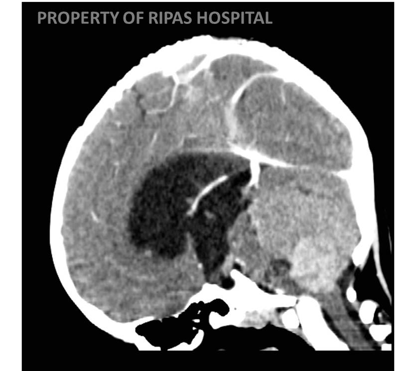
The usually presentation is with symptoms related to hydrocephalus.
RADIOLOGY
MRI with the administration of IV gadolinium is the diagnostic test of choice for medulloblastoma, although a CT has usually been performed first, following presentation.
A medulloblastoma is a midline vermilian mass, that usually displaces and so compress the 4th ventricle resulting in hydrocephalus. It demonstrates homogenous, avid contrast enhancement is typical. Leptomeningeal dissemination often termed ‘drop metastases’ are one of the potential complications of medulloblastoma following dissemination within the CSF, requiring MR imaging for diagnosis.
|
|
Location the key: (midline = medulloblastoma, hemispheric = pilocystic astrocytoma, arising in 4th ventricle = ependymoma and anterior to the 4th ventricle = brainstem glioma)
Images prepared by Dr Ian Bickle, Consultant Radiologist, RIPAS Hospital, Brunei Darussalam.
All images are copyrighted and property of RIPAS Hospital.
![]()