IMAGE OF THE WEEK
WEEK 15
|
|
|
PNEUMOPERITONEUM
Pneumoperitoneum is a common surgical presentation, due largely to a perforated viscus. The common sites of perforation are the sigmoid colon and dudodenum.
The query of perforation in the first instance merits an erect plain radiograph of the chest and is normally accompanied by an abdominal radiograph, acquired as usual in the supine position. The chest radiograph is by far the more sensitive of the two films, with free gas below the diaphragm confirming the diagnosis of perforation.
The abdominal radiograph is more difficult to assess, as the free gas within the peritoneal cavity is diffusely distributed and largely positioned anteriorly. However, there are a number of well described signs on plain abdominal radiograph.
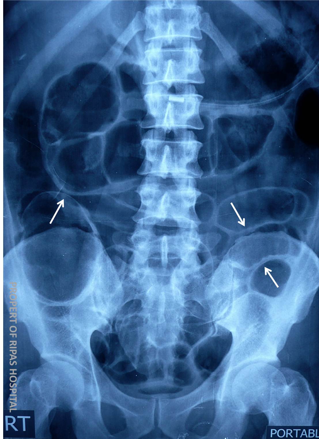
The most frequently identified is Rigler’s sign (a.k.a. The double wall sign - white arrows), when the bowel wall is crispy identified, almost giving a 3D appearance to the bowel, as there is gas on either side of the bowel wall. Gas as normal within the lumen of the bowel and gas outside the bowel wall (within the peritoneum cavity).
Other less commonly identified plain films findings of pneumoperitoneum include:
The falciform ligament sign (falciform ligament outline due to gas within the peritoneal cavity)
Triangles of gas (free gas trapped between 3 bowel loops)
The continuous diaphragm sign
The’ football’ sign
Images prepared by Dr Ian Bickle, Consultant Radiologist, RIPAS Hospital, Brunei Darussalam.
All images are copyrighted and property of RIPAS Hospital.
![]()