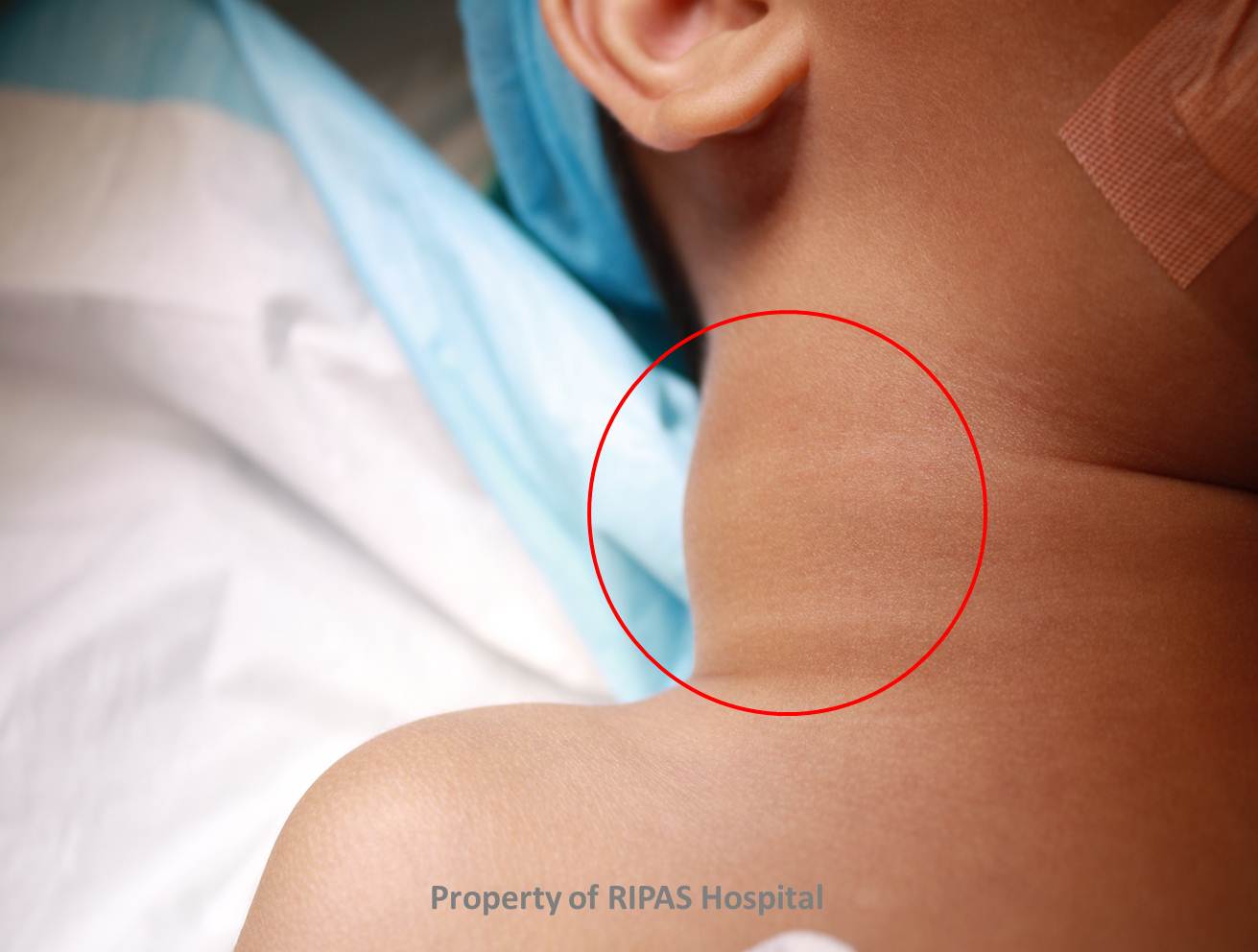
Figure 1: Cystic Hygroma of the neck (Click on picture to enlarge)
IMAGE OF THE WEEK 2012
WEEK 15
CYSTIC HYGROMA
|
|
Figure 1: Cystic Hygroma of the neck (Click on picture to enlarge) |
|
Cystic hygroma (CH) is an abnormal collection of lymphatic channels, which does not drain into the normal lymphatic system, thus forming a cytic lesion. It can be found in any anatomic subsite in the body but most commonly are found in the posterior triangle of the neck, with a left-sided predilection (75%). Other sites affected are axilla in 20%, infrequently in the mediastinum, groin and retroperitoneum. It is also known as cystic lymphangioma or macrocytic lymphatic malformation and was first described by Redenbacker in 1828.
CCHs or lymphagiomas are thought to arise from a failure of the lymphatic channels to connect and drain into the venous system with abnormal budding of lymphatic tissue and sequestration of lymphatic rests that retain their embryonic growth potential. The difficulty about managing this condition is the tendency of the lymphatic channels to infiltrate into adjacent structures or dissect along fascial planes.
There are 3 types of CHs, capillary, cavernous or cystic, depending on the nature of the surrounding tissue. CHs in the neck as is in the case above, tend to form in loose areolar tissue, whereas capillary and cavernous forms of lymphangiomas then to arise in muscle tissue. CHs can be congenital or acquired, the latter arising from trauma including surgery, inflammation or obstruction of lymphatic drainage. Studies have shown that lymphangiomas then to enlarge due to engorgement rather than cellular proliferation. With the case above, the CH enlarged and became visible after a fall on the right side of the neck resulting in haemorrhage into the cystic spaces of the CH. Molecular studies suggested that vascular endothelial growth factor C (VEGF-C) and its receptors may play an important role in the development of lymphatic malformations.
Incidence of CH is estimated at 1:6000-16000 live births. Mortality has been reported as high as 2-6% secondary to pneumonia, bronchiectasis and airway compromise, usually associated with large CH lesions. No racial or gender predominance have been reported. Most CHs (50-65%) are evident at birth, 80-90% presenting by 2 years of age.
Clinical presentation of CH vary depending on the location. Most are asymptomatic and become visible particularly in the neck following sudden cystic enlargement either from haemorrhage or infection. Compromisation of airway from large CHs results in stridor or obstructive sleep apnea syndrome (OSAS). There may be feeding difficulties resulting in failure to thrive.
CHs are typical soft, painless and compressible. Because of the cystic nature, they typically transilluminate well.
Imaging is the best modality for investigating these lesions with ultrasound being the first line modality. CT and MRI will further delinate the cystic lesion better particularly the relationship of the lesion to underlying structure and are particularly useful in mediastinal CHs, for which ultrasound is not suitable.
Staging of CHs
|
Giguere’s categorization of lymphangiomas |
|
|
Macrocystic |
Cystic spaces at least 2cm |
|
Microcystic |
Spaces less than 2cm |
|
Mixed lesions |
|
|
|
|
|
De Serres’s Staging for CH |
|
|
Stage I |
Unilateral infrahyoid (17% complication rate) |
|
Stage II |
Unilateral suprahyoid (41% complication rate) |
|
Stage III |
Unilateral and both infrahyoid and suprahyoid (67% complication rate) |
|
Stage IV |
Bilateral suprahyoid (80% complication rate) |
|
Stage V |
Bilateral infrahyoid and suprahyoid (100% complication rate) |
Medical management of CHs include watchful waiting or administration of sclerosing agents including OK-432, bleomycin, pure ethanol, sodium tetradecyl sulfate and doxycycline.
The mainstay of treatment is complete surgical excision. Unless the margins are completedly excised, recurrence can occur. Because of its close and infiltration into surrounding tissue, complete excision is difficult and is achieved in only 40% of cases. This is true of the microcystic type lesions because of their intimate association with surrounding tissues. Non surgical technique of removing these lesions include laser therapy which is a recent advances. Radiofrequency ablation has been advocated for use with intraoral lesions.
Image and text contributed and prepared by
Mr William Chong, Department of Surgery, RIPAS Hospital, Brunei Darussalam.
All images are copyrighted and property of RIPAS Hospital.
![]()