IMAGE OF THE WEEK 2013
WEEK 16
CARDIOVOCAL SYNDROME (ORTNER'S
SYNDROME)
|
 |
 |
|
|
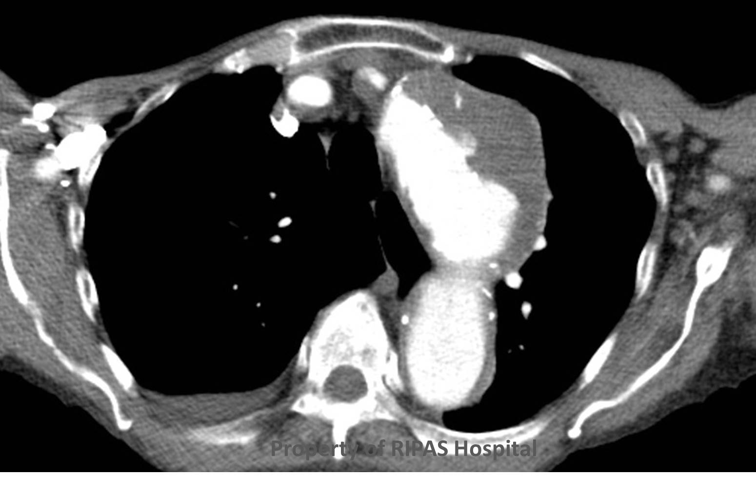
Figure 1a: Aortic arch aneurysm with traction injury
to the left phrenic nerve resulting in vocal cord palsy, a condition
known as Cardiovocal syndrome or Ortner's syndrome.
(Click on image to
enlarge) |
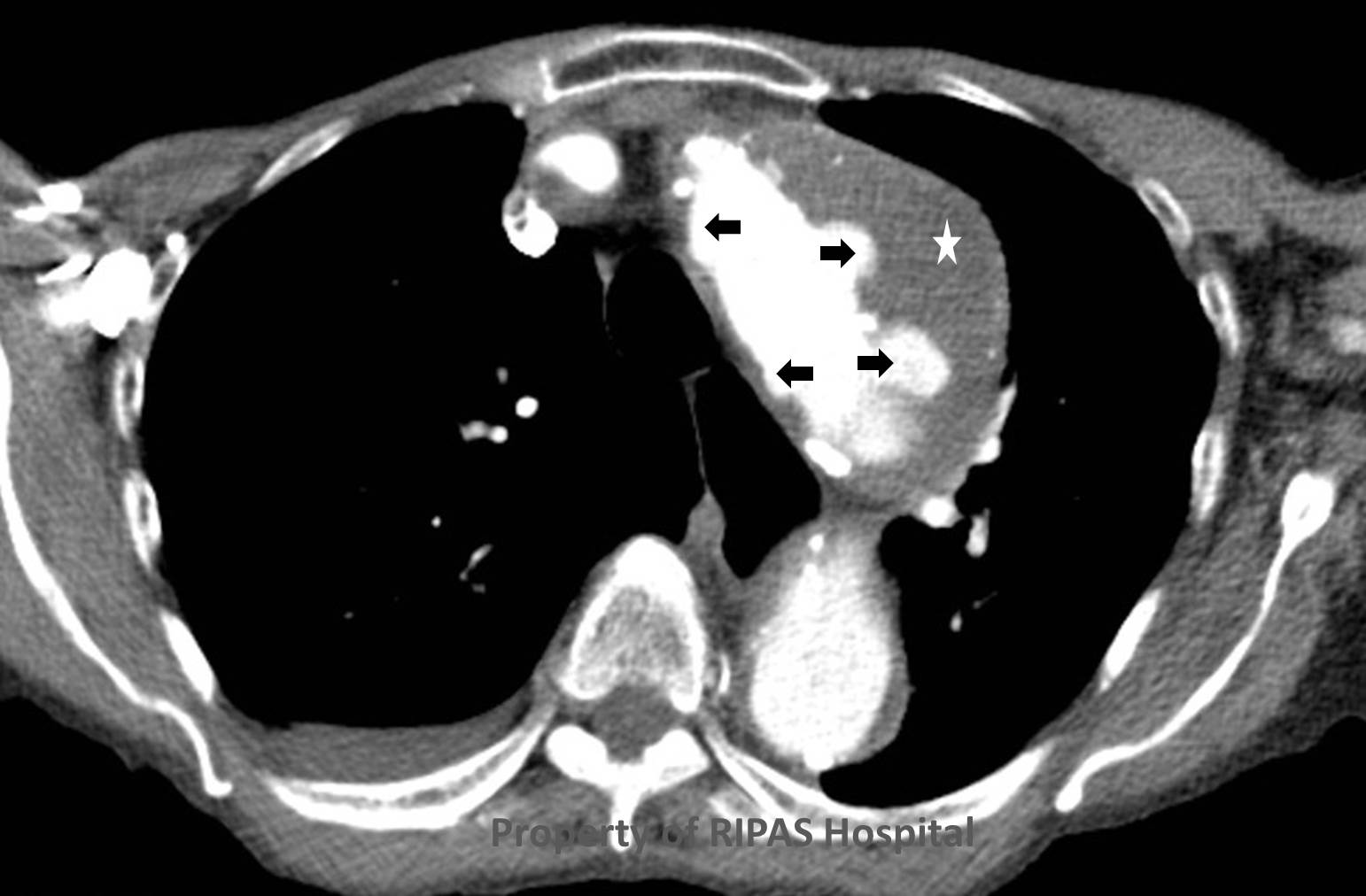
Figure 2: Aortic arch aneurysm showing mural ulcers
(black arrows) and intramural thrombus (white star).
(Click on image to
enlarge) |
|
|
|
|
|
Ortner's syndrome is a rare cardiovocal
syndrome
in which the a
recurrent laryngeal nerve
palsy
results from compression or traction injury in
cardiovascular
disease.
It is named after the author who described it in 1897 – Dr Ortner.
It is almost exclusively on the left side due to the long course of the left
recurrent laryngeal nerve around the aortic arch.
Ortner’s original description
was attributed to a case of left vocal fold paralysis due to compression of the
recurrent laryngeal nerve by a dilated left atrium in a patient with mitral
valve stenosis. Subsequent descriptions have been documented regarding other
intra-thoracic cardiac causes for cardio-vocal syndrome, including aortic arch
aneurysms, which cause traction type injury to the nerve, as in this case.
This elderly patient’s left vocal cord paralysis was found to be due to
compression from a large thoracic aortic aneurysm, involving the arch of aorta
(Figure 1). The aneurysm contains a large volume of atheroma and penetrating
ulcers (Figure 2 and 3).
|
 |
 |
|
Figure 3: Coronal plane CT chest showing the mural
ulcer (black arrow) and mural clot (white star) in the aortic arch
aneurysm.
(Click on image to
enlarge) |
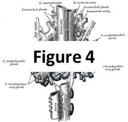
Figure 4: Schematic diagram showing the anatomical
pathway of the nerve
(Click on image to
access image) |
The left recurrent laryngeal nerve is not observed on CT. However, the inference
between the clinical and radiological findings, with knowledge of the anatomical
path of the nerve (Figure 4 – courtesy of Wikipedia) and absence of a cause for
the palsy in the head and neck, clinches the diagnosis.
Images and text contributed by
Dr Ian Bickle, Department of Radiology,RIPAS Hospital
All
images are copyrighted and property of RIPAS Hospital.

