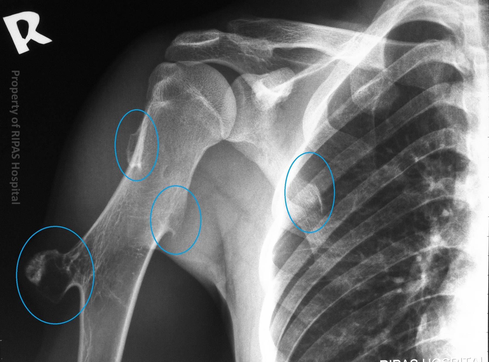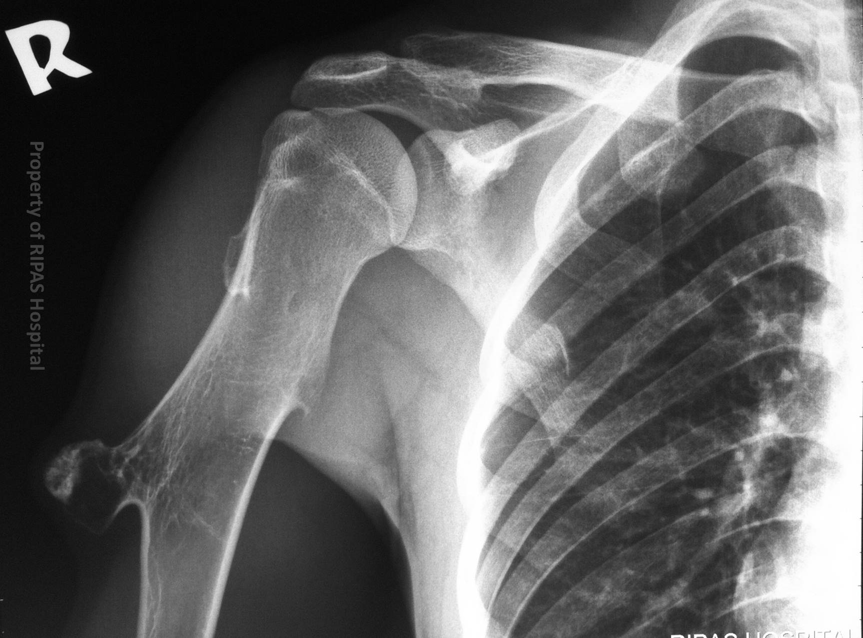
Figure 1: Plain radiograph of the right shoulder, showing multiple osteochondromas (exostoses) at mid humeral shaft.
(Click on image to enlarge)
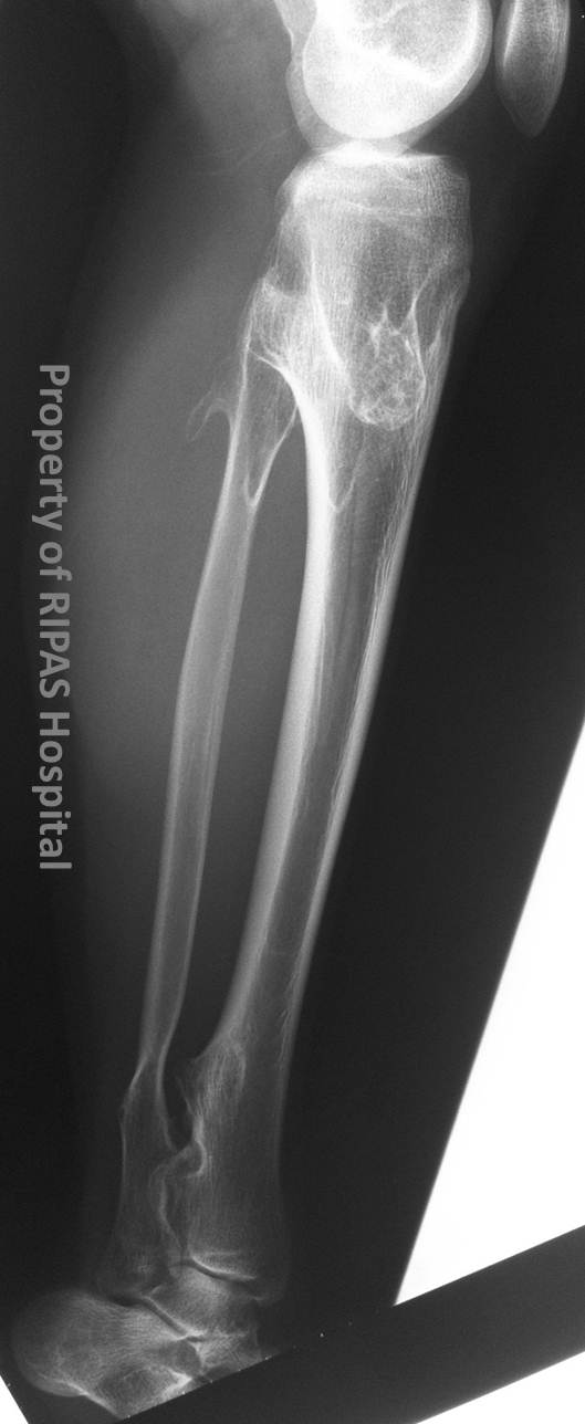
Figure 2: Plain radiograph of right forearm showing multiple exostoses.
(Click on image to enlarge)
IMAGE OF THE WEEK 2013
WEEK 18
Diaphyseal achalasia ( Multiple Hereditary Exostoses )
|
|
|
|
|
Figure 1: Plain radiograph of the right shoulder, showing multiple osteochondromas (exostoses) at mid humeral shaft. (Click on image to enlarge) |
Figure 2: Plain radiograph of right forearm showing multiple exostoses. (Click on image to enlarge) |
|
Diaphyseal achasia is a condition characterised by development of multiple osteochondromas (exostoses) throughout, but not exclusively involving, the appendicular skeleton (Figures 1 and 2). Osteochondromas are developmental anomalies, although typically classified as benign bone tumours. Even the term benign is misleading, with a small number (around 1%) undergoing malignant transformation. However, this figure is significantly larger in those with diaphyseal achalasia, given the number of osteochondromas is high (Figure 3).
|
|
|
Figure 3: Annotated image of Figure 1 showing the multiple osteochondromas circled by the blue ring. Click on image to enlarge. |
As the name implies these lesions arise from the diaphysis of the bone, most commonly around the knee and shoulder. The lesions almost always point away from the joint.
Malignant transformation actually occurs in the cartilage cap, for which MRI is the modality of choice assess, as this is not visualised on plain radiograph.
This is just one of several potential complications from these lesions.
The others include:
1. Fracture – usually through the neck and not infrequently the reason for first being identified.
2. Neurovascular impingement
3. Cosmetic deformity with associated pain
4. Bursitis
Images and text contributed by
Dr Ian Bickle, Department of Radiology,RIPAS Hospital
All images are copyrighted and property of RIPAS Hospital.
![]()