Figure 1a
Figure 1b
IMAGE OF THE WEEK
WEEK 19
|
|
|
|
|
Figure 1a |
Figure 1b |
SUBARACHNOID HAEMORRHAGE
Subarachnoid Haemorrhage (SAH) is bleeding into the subarachnoid space - area between the arachnoid membrane and the pia mater surrounding the brain. Subarachnoid haemorrhage may occur spontaneously, usually as a result of a ruptured cerebral aneurysm, or may occur due to a head injury. The latter is termed as traumatic subarachnoid haemorrhage.
Three quarters of spontaneous subarachnoid haemorrhages are due to an underlying cerebral (‘Berry’) aneurysms. Berry aneurysms are associated with other conditions, such as Marfan’s disease, Ehler Danlos and Adult Polycystic Kidney Disease. Of the remaining causes arteriovenous (AV) malformations are the commonest other cause (10%). Rarer causes include; eclampsia, underlying blood dyscrasias and hypertension. Seven (7%) of all strokes are caused by SAH from ruptured berry aneurysms.
Patient presenting with a subarachnoid haemorrhage classically complained of a sudden onset ‘thunderclap’ occipital headache, which is unremitting.
|
|
 |
|
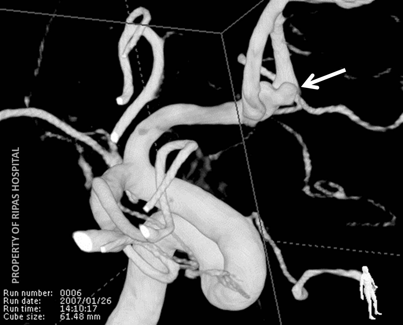
Figure 2a |
Figure 2b |
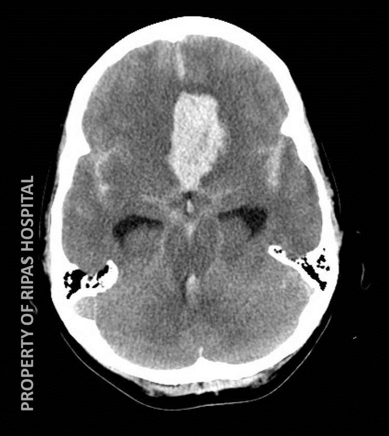
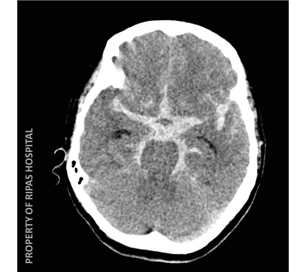
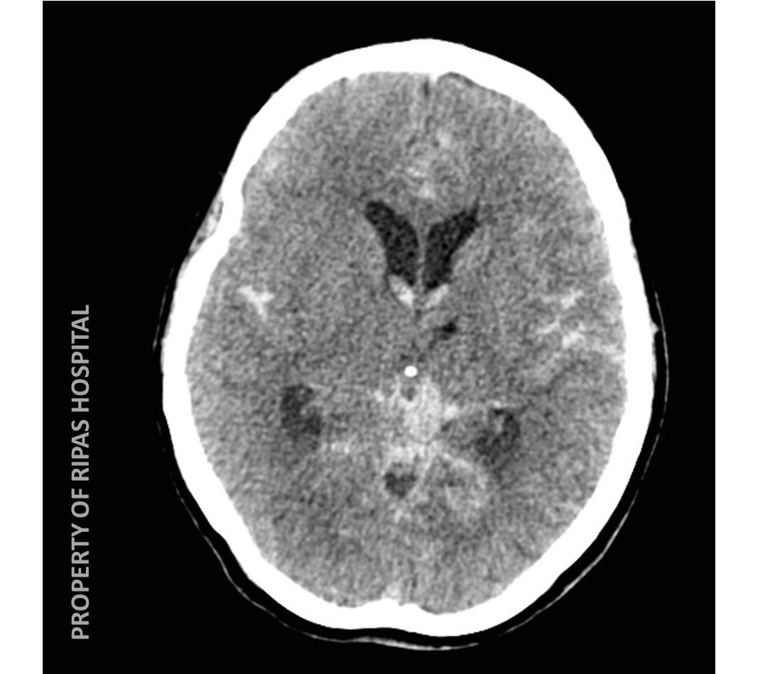
Urgent CT is the first line investigation, which will identify acute haemorrhage within the subarachnoid space, typically within the basal cisterns, sulci and fissures of the cerebral hemispheres. Frequently blood extends into the ventricular system, which may result in obstructive hydrocephalus. The distribution of haemorrhage within the brain, may point towards the possible aneurysm site, for example an anterior parafalcine haematoma is highly suggestive of an anterior communicating artery (ACOM) aneurysm bleed (Figure 2a). CT cerebral angiography (Figure 2b) and or catheter cerebral angiography may be performed to identify an underlying aneurysm to allow for definitive treatment. This may take the form of traditional surgical ‘clipping’ or more commonly now in most centres endovascular coil embolisation.
|
|
 |
|
Figure 3a |
Figure 3b |
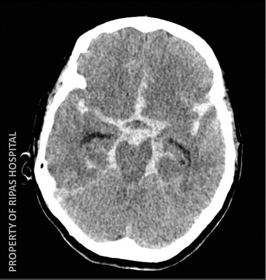
Extensive subarachnoid blood within the basal cisterns has been termed the ‘hanging chicken’ sign (Figure 3)
Images prepared by Dr Ian Bickle, Consultant Radiologist, Department of Radiology, RIPAS Hospital, Brunei Darussalam and edited by Mr CF Chong.
All images are copyrighted and property of RIPAS Hospital.
![]()