IMAGE OF THE WEEK 2013
WEEK 19
SPINAL SCHWANNOMAS
|
 |
 |
 |
|
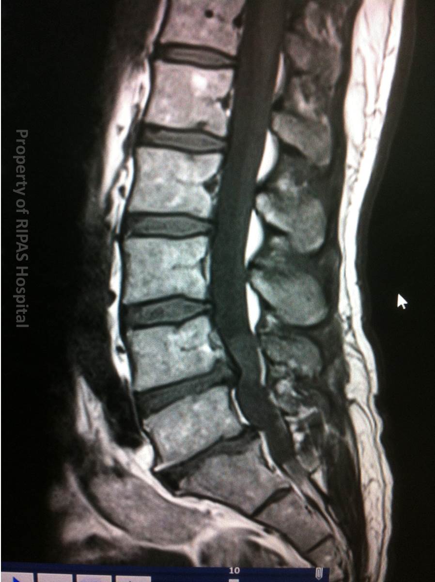
Figure 1: MRI T1 sequence saggital section of the
lumbar spine showing an isointense space occupying lesion at L5 level.
(Click on image to
enlarge) |
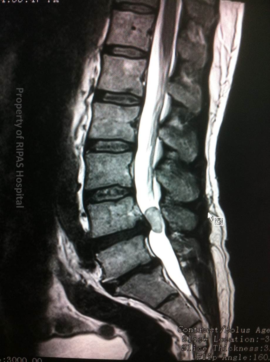
Figure 2: MRI T2 sequence saggital section of the
same lumbar spine showing a hyperintense space occupying lesion at L5
level.
(Click on image to
enlarge) |
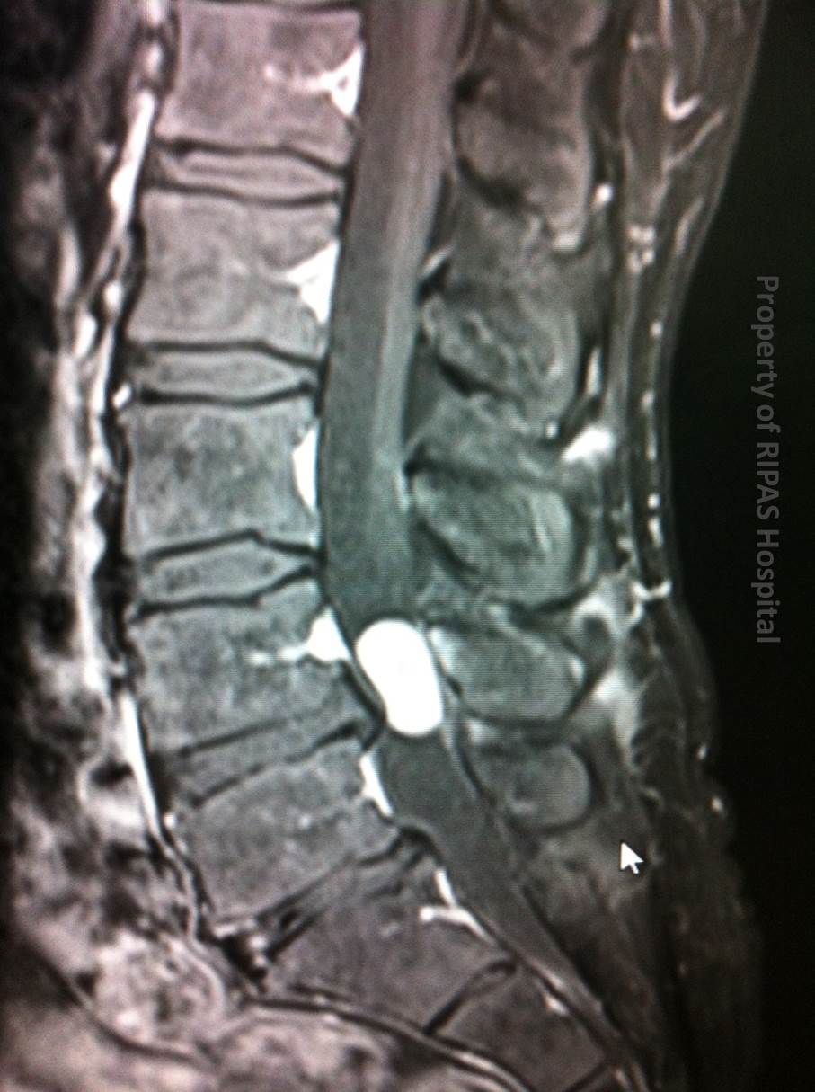
Figure 3: MRI saggital section of the same lumbar
spine following gadolinium administration, showing an avidly enhancing
space occupying lesion at L5 level.
(Click on image to
enlarge) |
|
|
|
|
Spinal schwannomas are
benign tumours arising from nerves in the spinal canal.
Schwannomas can arise from any nerves in the body, perhaps the
best known being the vestibular (aka acoustic) schwannomas arising at the
cerebellopontine angle in the
brain.
Tumours of the spinal canal are classified on their location with
respect to the dura and the cord. Spinal schwannomas fall into the intradural
extramedullary category, being within the thecal sac, but outside of the cord.
They are the commonest tumour in this category.
These lesions are usually very well defined arising from dorsal
nerve roots and present with pain or incidentally on imaging performed for an
alternative indication.
MRI is the only sensitive imaging modality for the detection of
schwannomas with the administration of contrast (gadolinium) greatly assisting
in their identification, especially if small.
A schwannoma typically has the following appearances on MRI:
-
Isointense on T1 (therefore hard to see on this sequence)
(Figure 1)
-
Hyperintense on T2 (Figure 2)
-
Avidly enhancing following gadolinium administration
(Figures 3 and 4)
Schwannomas and neurofibromas may be almost identical in
appearance, however schwannomas often have small amounts of cystic of
haemorrhagic change within, clinching the diagnosis.
Surgery is indicated if symptoms are problematic, as these
tumours are almost universally histopathologically benign.
|
 |
|
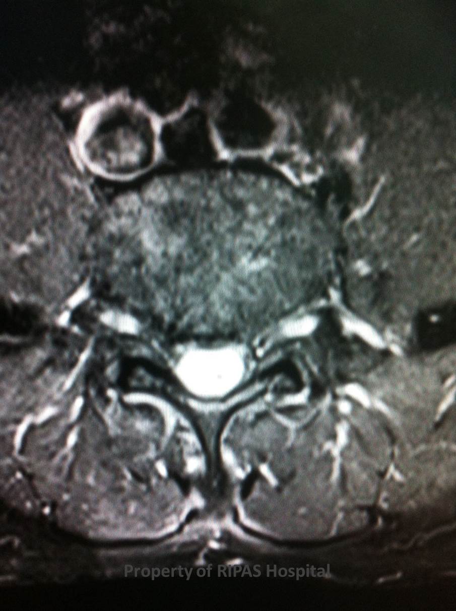
Figure 4:
MRI transverse section of the lumbar spine at L5
level following gadolinium administration, showing an avidly enhancing
space occupying lesion.
(Click on image to
enlarge) |
Images and text contributed by
Dr Ian Bickle, Department of Radiology,RIPAS Hospital
All
images are copyrighted and property of RIPAS Hospital.
