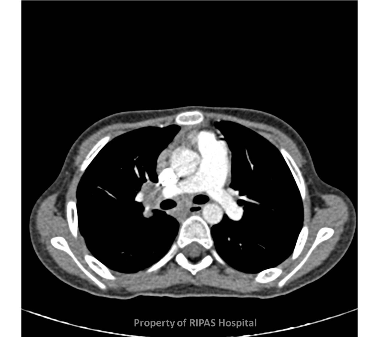
Figure 1: Extra-pulmonary involvement with mediastinal lymphadenopathy. (Click on picture to enlarge)
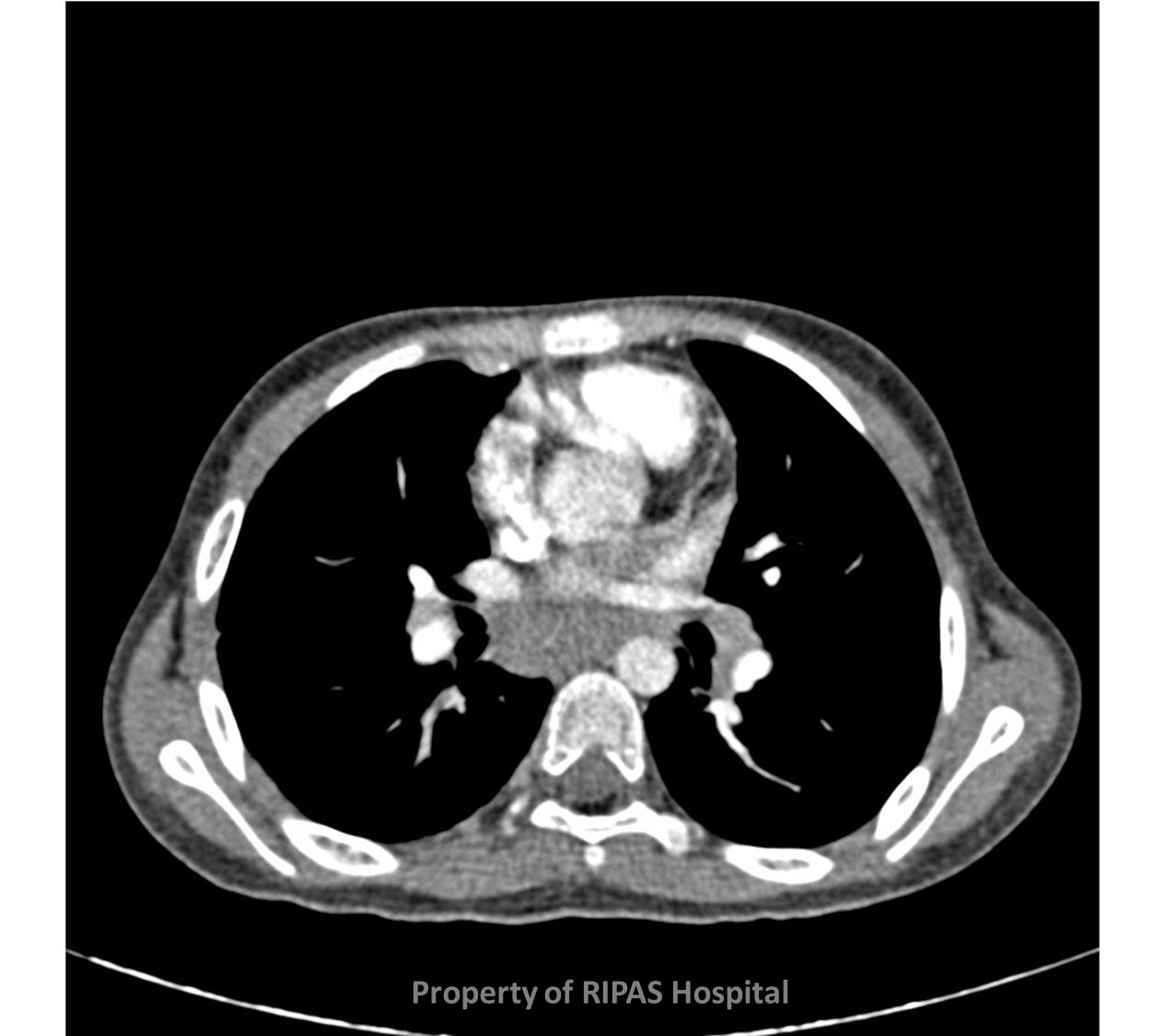
IMAGE OF THE WEEK 2012
WEEK 19
EXTRA-PULMONARY TUBERCULOUS: PERITONITIS
Although most commonly associated with the respiratory system tuberculosis (TB) is a true multi-system (Figures 1 & 2). Along with the genitourinary tract the gastrointestinal tract is one of the more common non-respiratory manifestations of TB. It however now occurs more commonly in isolation than in conjunction with positive chest x-ray findings (Figures 1 & 2). It is more common in those with underlying immunosuppression.
|
|
Figure 1: Extra-pulmonary involvement with mediastinal lymphadenopathy. (Click on picture to enlarge) |
|
|
Figure 2: Extra-pulmonary involvement with enlarged mediastinal lymphadenopathy (Click on picture to enlarge) |
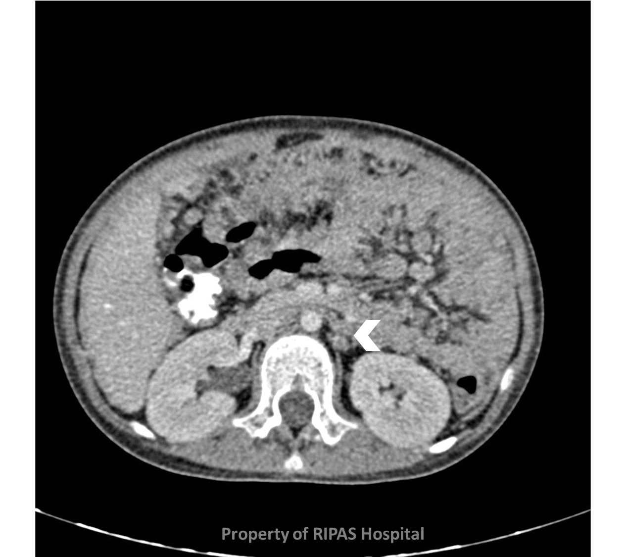
The Ileo-caecal area (white arrow) is by far the most commonly involved part of the gastrointestinal tract (Figures 3 & 4) with circumferential thickening of the bowel wall characteristic. There may be associated lymphadenopathy (White arrow head). The main differential diagnosis is lymphoma.
|
|
Figure 3: Gastrointestinal tract involvement with circumferential thickening of the bowel wall and lymphadenopathy as indicated by the white arrow head and matting of bowel. (Click on picture to enlarge) |
|
|
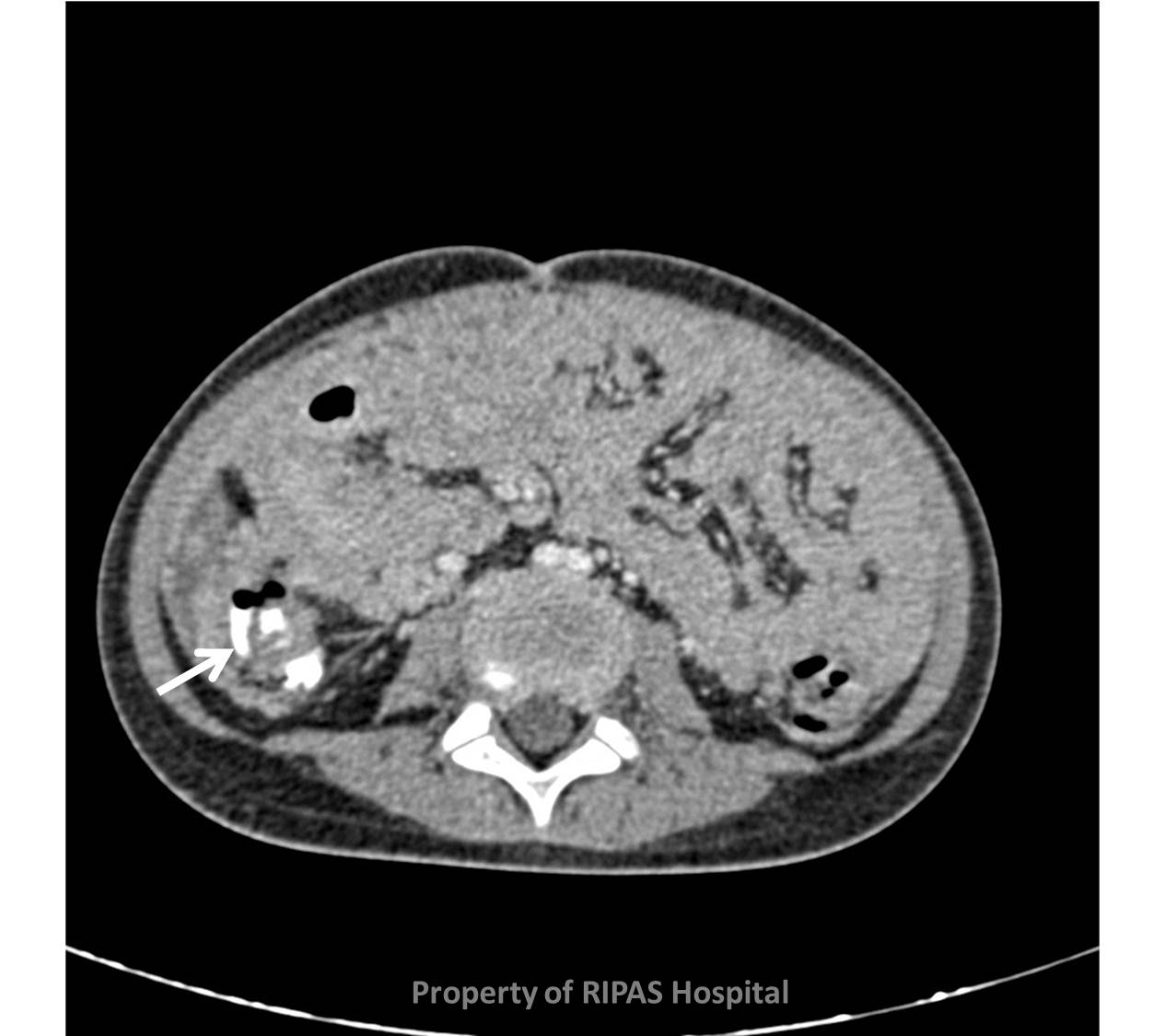
Figure 4: Ileo-caecal valve involvement as indicated by white arrow with matting of bowel. (Click on picture to enlarge) |
Peritoneal involvement is much less common, and can be either ‘dry’ or ‘wet’ type. Typically there is thick peritoneal soft tissue (or ‘cake’), which appears almost identical to diffuse peritoneal malignancy (Figure 3 & 4). This may be associated with large volume ascites (wet type) or none (dry type). Other features include mesenteric lymphadenopathy and ‘matting’ of the bowel (Figure 5). The lymph nodes classically are conglomerate in nature with a low attenuation centre.
|
|
Figure 5: peritoneal involvement of 'dry' type with matting of bowel and thickened bowel wall. (Click on picture to enlarge) |
Images and text contributed and prepared by
Dr Ian Bickle, Department of Radiology,
All images are copyrighted and property of RIPAS Hospital.
![]()