IMAGE OF THE WEEK
WEEK 2
COLORECTAL CARCINOMA
One of the commonest malignancies in Brunei, like most developed nations.
The majority are adenocarcinomas.
Unlike many other primary tumours partial liver resection considered for solitary metastasis.
Radiographic Features
 |
 |
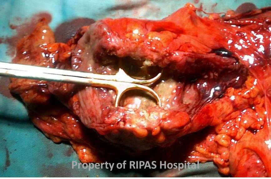
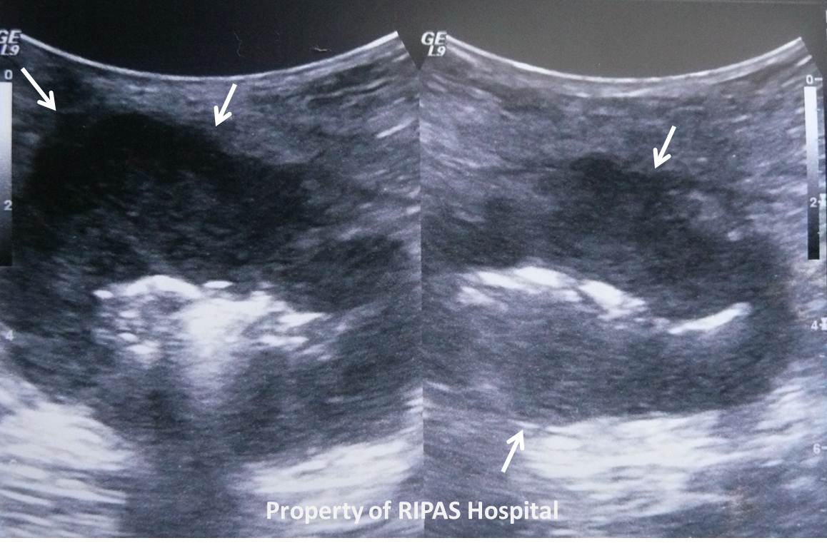
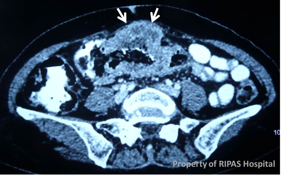
In this case on both the ultrasound and CT a circumferentially thickened transverse colon, with extra-serosal tumour extension, is apparent.
Common sites of metastases are loco-regional (pericolic) nodes and the liver.
Rectal malignancy considered separately in terms of imaging with ideally pre-operative MRI as well as staging CT.
|
TNM (Tumor, Node, Metastasis) Staging System for Colorectal Cancer |
|
|
Tumor Stage |
Description |
|
T1 |
Tumor invades submucosa. |
|
T2 |
Tumor invades muscularis propria. |
|
T3 |
Tumor invades through the muscularis propria into the subserosa, or into the pericolic or perirectal tissues. |
|
T4 |
Tumor directly invades other organs or structures, and/or perforates. |
|
|
|
|
Node |
Description |
|
N0 |
No regional lymph node metastasis. |
|
N1 |
Metastasis in 1 to 3 regional lymph nodes. |
|
N2 |
Metastasis in 4 or more regional lymph nodes. |
|
|
|
|
Metastasis |
Description |
|
M0 |
No distant metastasis. |
|
M1 |
Distant metastasis present. |
Prepared by Dr Ian Bickle, Consultant Radiologist, RIPAS Hospital, Brunei Darussalam.
All images are copyrighted and property of RIPAS Hospital.