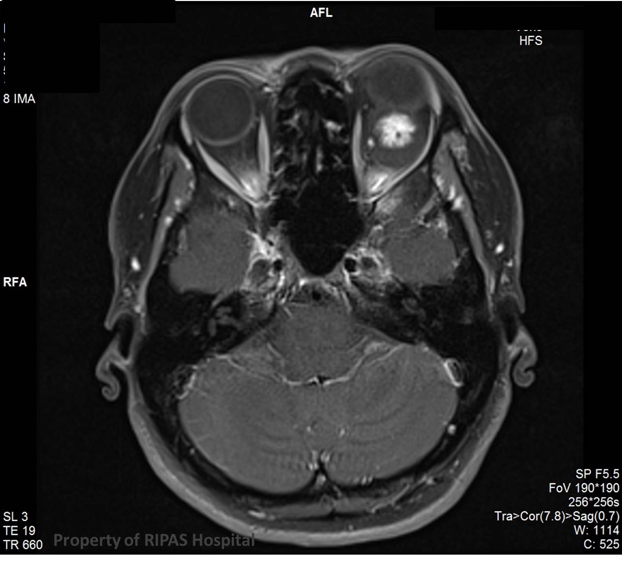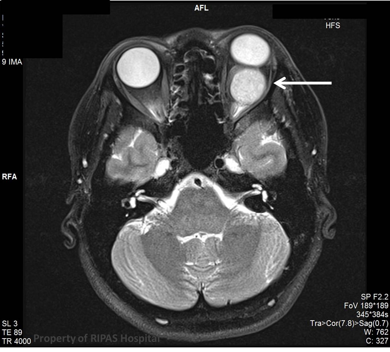
Figure 1a: Optic cavernous haemangioma appearing as a solid white mass behind the left eye ball as indicated by the white arrow. (Click to enlarge picture)
IMAGE OF THE WEEK 2012
WEEK 21
OPTIC CAVERNOUS HAEMANGIOMA
|
|
Figure 1a: Optic cavernous haemangioma appearing as a solid white mass behind the left eye ball as indicated by the white arrow. (Click to enlarge picture) |
A cavernous haemangioma (Figure 1a) of the orbit is the commonest vascular lesion of the orbit in adults, characterized by endothelial lined cavernous spaces. It is not a ‘true haemangioma’.
It typically presents in middle age (30 - 50 years of age) and with a slight female predilection.
Clinical presentation is usually with a slowly growing orbital mass resulting in proptosis. Diplopia and visual field defects (from optic nerve compression) can also occur.
Cavernous haemangiomas are well circumscribed masses with a thin fibrous pseudocapsule, without prominent arterial supply – the reason why these lesions show a relatively slow, sometimes initially patchy enhancement, that on delayed phases fills in. As flow is slow and vascular spaces large, areas of thrombosis can occur.
Although cavernous haemangiomas can be located anywhere within the orbit the vast majority (more than 80%) are in the intra-conal space. They are nearly always round or oval and although they may abut the globe (as in this case), they do no deform or infiltrate it
MRI is the most sensitive imaging modality, although CT may have taken place first.
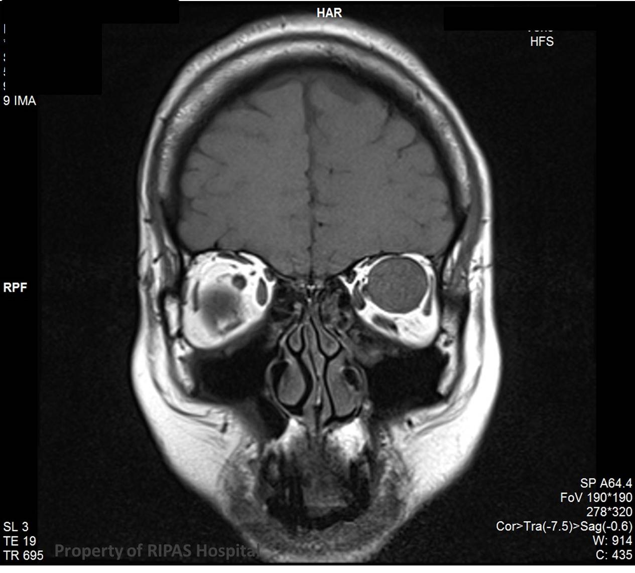
The mass is isointense to muscle on T1 (Figure 1b)
|
|
Figure 1b: Optic cavernous haemangioma appearing as a solid white mass behind the left eye ball as indicated by the white arrow. (Click to enlarge picture) |
Hyper-intense to muscle on T2 with a thin pseudocapsule is of low intensity (Figure 2)
|
|
Figure 2: (Click to enlarge picture) |
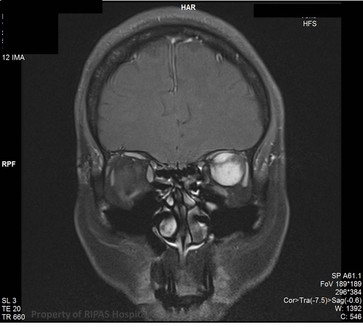
T1 contrast : slow heterogeneous patchy enhancement with delayed homogenous filling (Figure 3).
|
|
Figure 3: (Click to enlarge picture) |
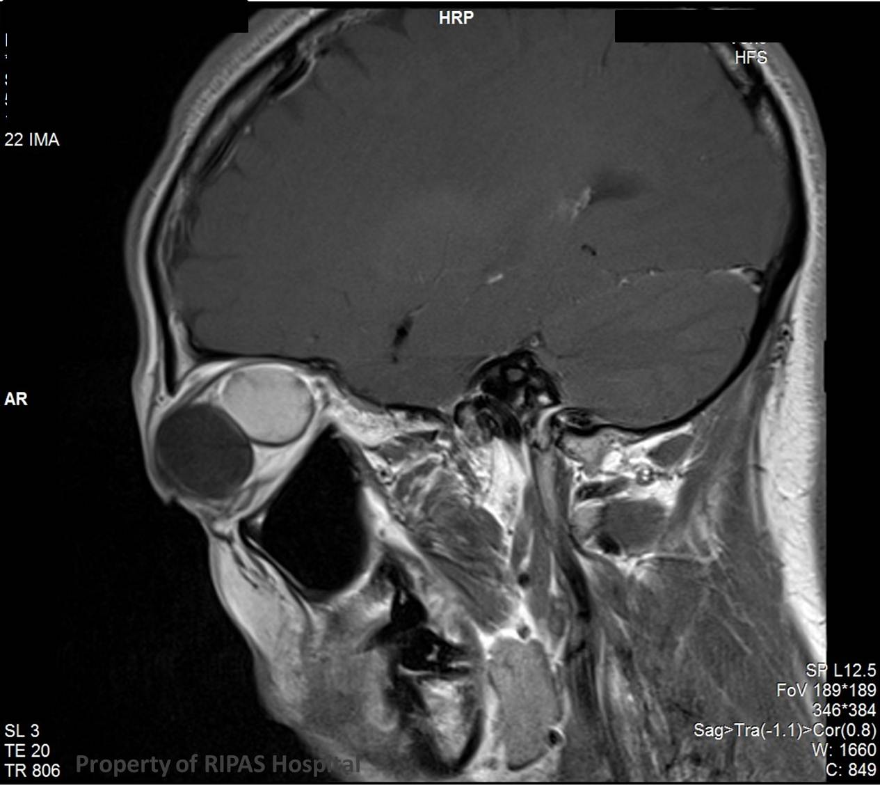
Visual deterioration may occur clinically due to mass effect on the optic nerve (Figure 4).
|
|
Figure 4: (Click to enlarge picture) |
Images and text contributed and prepared by
Dr Ian Bickle, Department of Radiology,
All images are copyrighted and property of RIPAS Hospital.
![]()