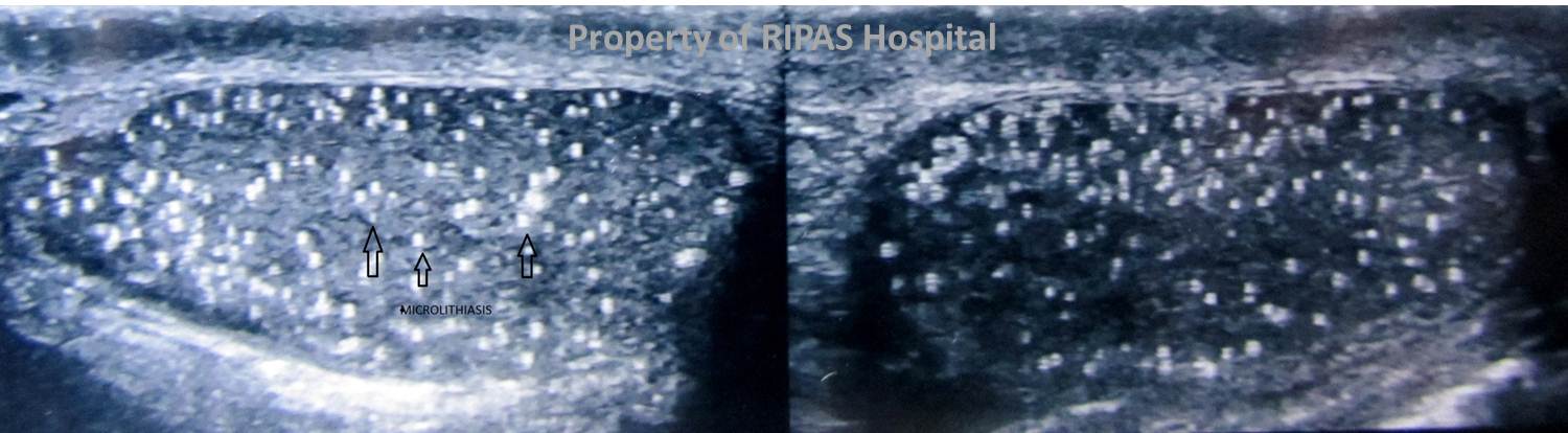
Figure 1: . Testicular microliathiasis as shown by the white calcified particles in the substance of the testes indicated by the arrows. (Click to enlarge picture)
IMAGE OF THE WEEK 2012
WEEK 22
TESTICULAR MICROLITHIASIS
|
|
|
|
Figure 1: . Testicular microliathiasis as shown by the white calcified particles in the substance of the testes indicated by the arrows. (Click to enlarge picture) |
Testicular microlithiasis is the deposition of small amounts of calcification with the testicles. It is identified incidentally in up to 1% of men undergoing scrotal ultrasound.
It is also present in up to 50% of males with a germ cell testicular tumour; however it is very common in patients without cancer. It remains debatable as to whether a direct relationship exists between microlithiasis and testicular malignancy and if those with microlithiasis should have regular surveillance ultrasound imaging.
Microlithiasis is associated with numerous conditions including: Testicular germ cell tumour, Kleinfelter syndrome, Cryptorchidism, Testicular infarct, Down syndrome.
The gold standard imaging modality for assessment of the testicle is ultrasound. In limited cases there is a role for MRI. On ultrasound microlithiasis appears as tiny hyperechoic foci ranging in diameter from 1 to 3 mm, without acoustic shadowing. The foci are typically scattered in the testicular parenchyma and may be distributed peripherally, segmentally or diffusely.
Images and text contributed and prepared by
Dr Ian Bickle, Department of Radiology,
All images are copyrighted and property of RIPAS Hospital.
![]()