IMAGE OF THE WEEK 2012
WEEK 24
Leptomeningeal metastases
|
 |
 |
|
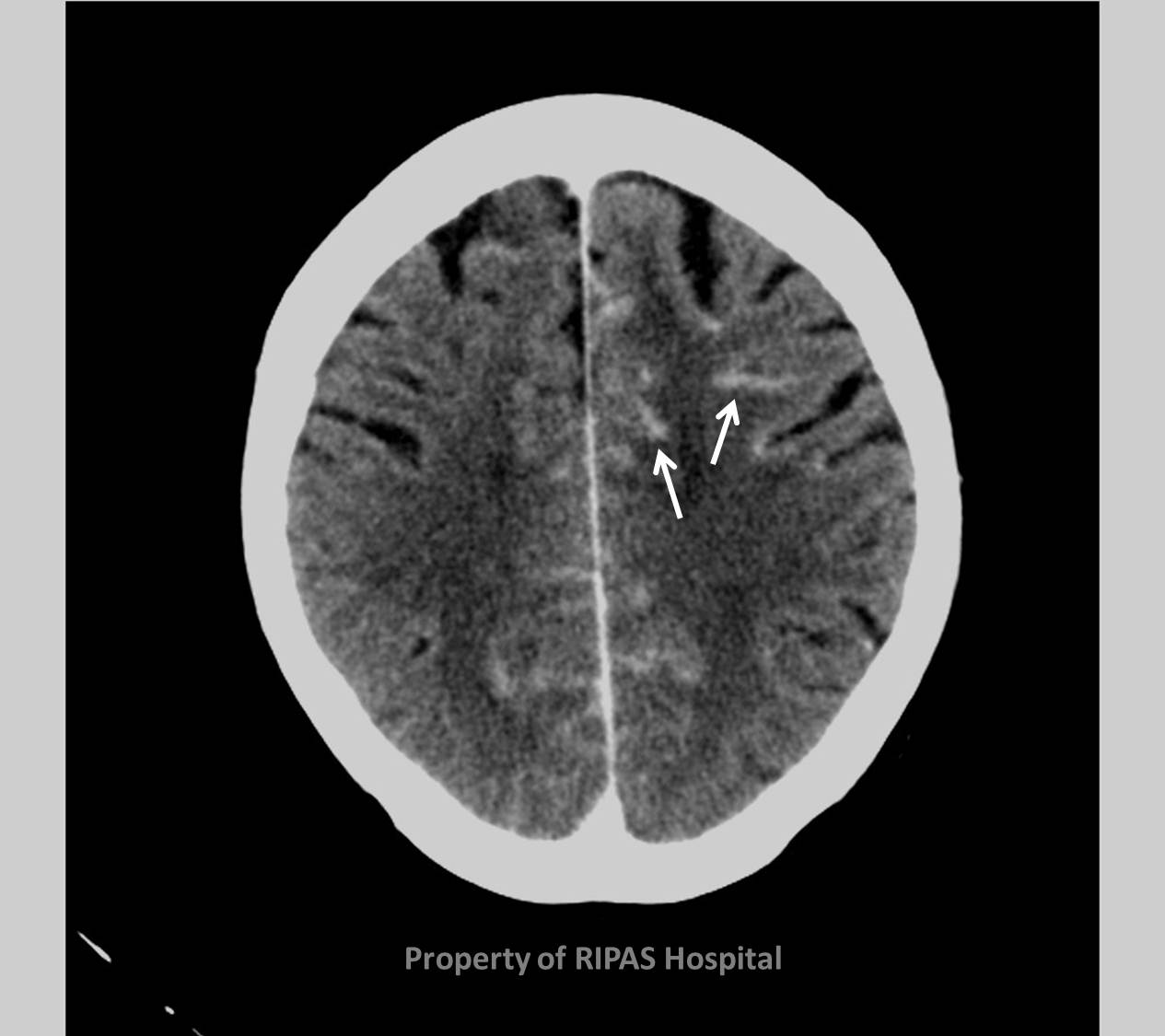
Figure 1: CT scan of the brain showing contrast
enhancement of the leptomeninges as "icing sugar coating", indicated by
the white arrows.
(Click to
enlarge picture) |
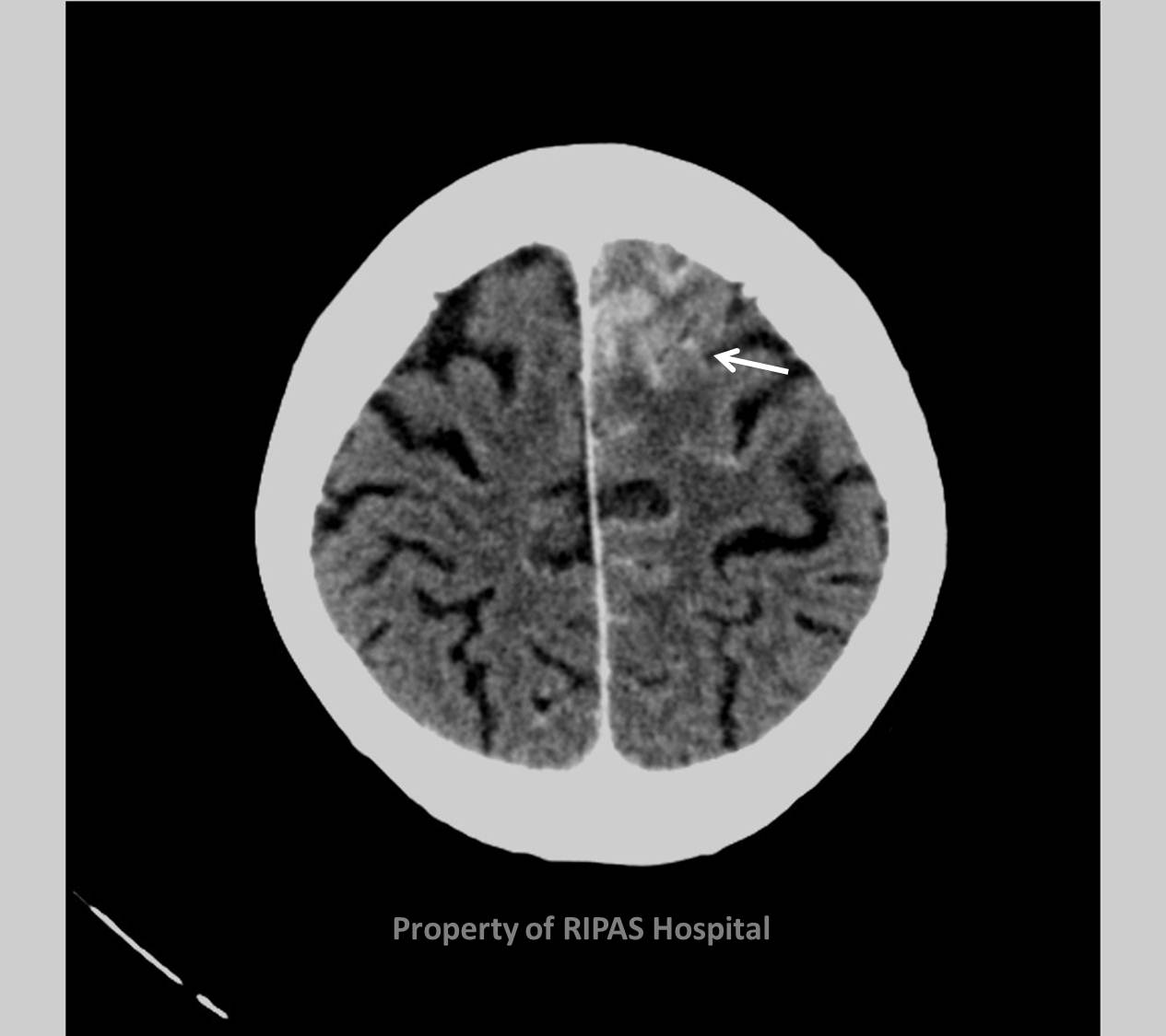
Figure 2: CT scan of the brain showing contrast
enhancement of the leptomeninges as "icing sugar coating", indicated by
the white arrows. (Click to
enlarge picture) |
Leptomeningeal metastases are the spread of malignant cells through the
cerebrospinal fluid – this can be either from a primary CNS tumour or more
commonly from a distant primary tumour. The metastatic deposits may be within
the brain or ‘coated’ along the leptomeninges of the spinal cord.
The clinical presentation may be quite non-specific, such as headache or being
‘out of sorts’.
Leptomeningeal metastases are best and often only identified after a post
contrast imaging study – either CT or MRI. The latter is most sensitive, most
commonly appreciated as linear enhancement over the leptomeninges in an
‘icing sugar coating’, like that over the top of a cake (Figures 1 & 2).
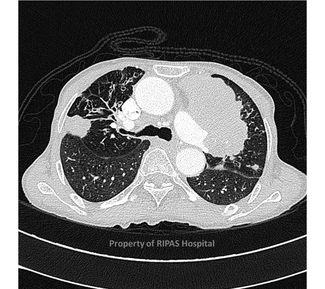
The commonest primary tumours associated with leptomeningeal metastases are lung
and breast cancer, but can occur in other tumours, such as melanoma (Figure 3).
|
 |
|
|
Figure 3: CT scan of the chest showing a left T4
NSCLC with a right middle lobe metastasis. (Click to
enlarge picture) |
|
Images and text contributed and prepared by
Dr Ian Bickle, Department of Radiology,
All
images are copyrighted and property of RIPAS Hospital.
