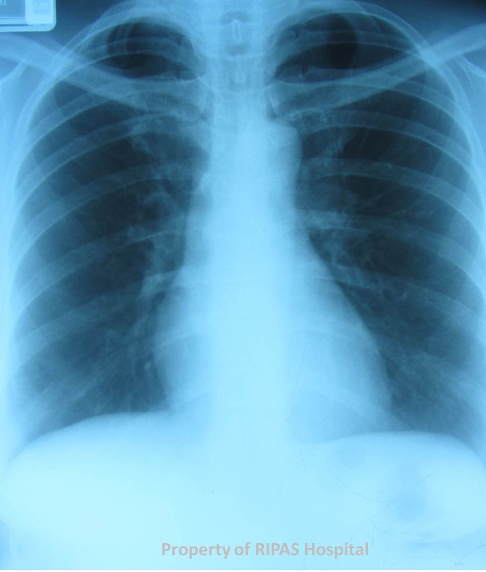
Figure 1: Chest X-ray showing a right cervical rib.
(Click to enlarge picture)
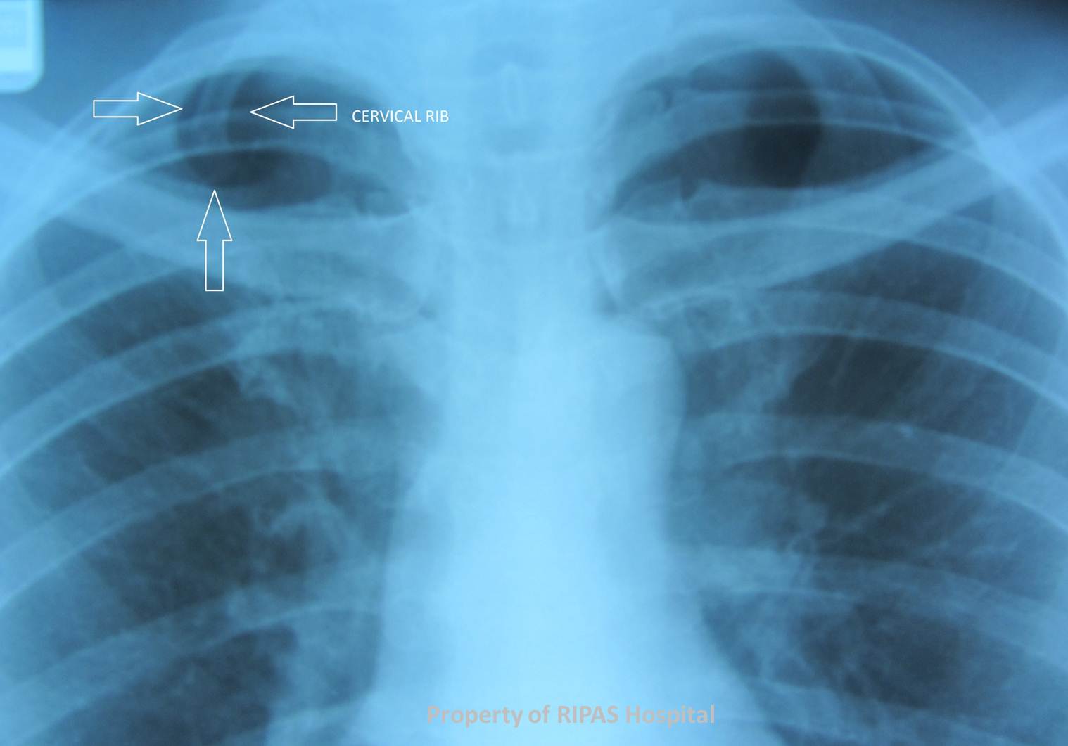
Figure 2: Annotated Chest X-ray pointing at the cervical rib.
(Click to enlarge picture)
IMAGE OF THE WEEK 2012
WEEK 27
CERVICAL RIB
|
|
|
|
Figure 1: Chest X-ray showing a right cervical rib. (Click to enlarge picture) |
Figure 2: Annotated Chest X-ray pointing at the cervical rib. (Click to enlarge picture)
|
A cervical rib is an accessory rib arising from the C7 vertebral body, which is often an incidental finding on chest x-ray. It is a rare condition, occurring in less than 1% of the population. It can however, be highly significant clinically, with patients presenting with symptoms of neurovascular compromised due to compression on the neurovascular bundle. Cervical rib is one cause of ‘thoracic outlet syndrome’ is a result of direct impingement on the subclavian vessels and or brachial plexus.
Compression on the brachial plexus causing neurological symptoms, such as paraesthesia is more common than ischaemia due to vascular involvement. Neurogenic symptoms usually arise following neck trauma in 80% of patients with cervical ribs, either following work-related injuries or repetitive stress or road traffic accidents. In 20% of patients with cervical ribs, symptoms appeared spontaneously.
Indications for surgical resection of the accessory rib are disabling pain, paraesthesia and failure of conservative treatment. Surgery involves resection of the accessory rib with or without excising the first rib. Failure rate of surgical treatment is high in those with work-related injuries (42%) compared with those arising following RTA (26%) or spontaneously (18%). Surgical success rate is also higher if both cervical and first ribs are resected together.
Reference
Sanders RJ, Hammond SL. Management of cervical ribs and anomalous first ribs causing neurogenic thoracic outlet syndrome. J Vasc Surg. 2002;36(1):51-6.
Images and text contributed and prepared by
Dr Ian Bickle, Department of Radiology, and Mr Chee Fui Chong, Department of Surgery.
All images are copyrighted and property of RIPAS Hospital.
![]()