IMAGE OF THE WEEK 2012
WEEK 28
Duplication of the IVC
|
 |
 |
|
Figure 1: Abdominal CT scan at the level of the
kidneys showing two circular structures on either side of the slightly
calcified aorta, which are both IVCs (White arrows).
(Click to
enlarge picture) |
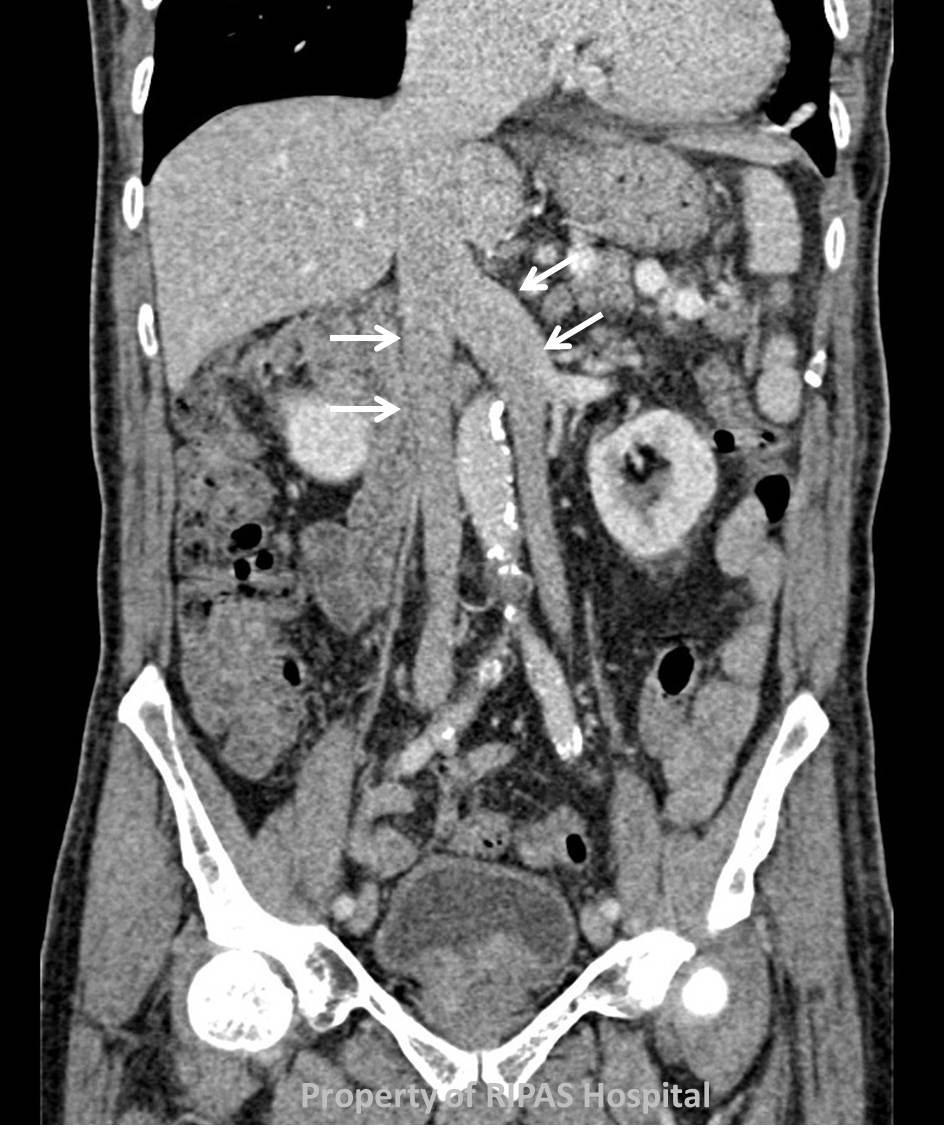
Figure 2: Coronal plane of the abdomen on CT scan at
the level of the IVC showing a duplicate IVC as indicated by the white
arrows.
(Click to
enlarge picture)
|
Various congenital variations of the inferior vena cava (IVC) have been
described. These include:
a.
Duplication of the IVC
b.
Azygous continuation of the IVC
c.
Left sided IVC
In duplication of the IVC, there are both left and right sided IVC’s inferior to
the renal veins, lying either side of the abdominal aorta. These continue on
either side to inform the common iliac vein. The left sided IVC usually ends at
the level of the left renal vein.
Although not seen in this case there may be asymmetry in size between the two
IVC’s.
Images and text contributed and prepared by
Dr Ian Bickle, Department of Radiology.
All
images are copyrighted and property of RIPAS Hospital.
