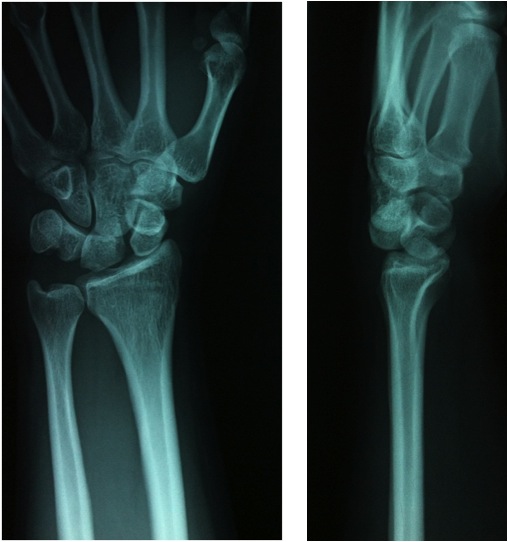
Figure 1: X-ray of the wrist
(AP and Lateral) showing perilunate dislocation
(Click on image to enlarge)
IMAGE OF THE WEEK 2013
WEEK 3
PERILUNATE DISLOCATION
|
|
|
|
Figure 1: X-ray of the wrist (Click on image to enlarge) |
Perilunate dislocations are potentially devastating wrist injuries that are not infrequently overlooked on initial imaging.
Perilunate dislocations typically occur in adults under 40 years of age with high impact hyperextension injury of the wrist.
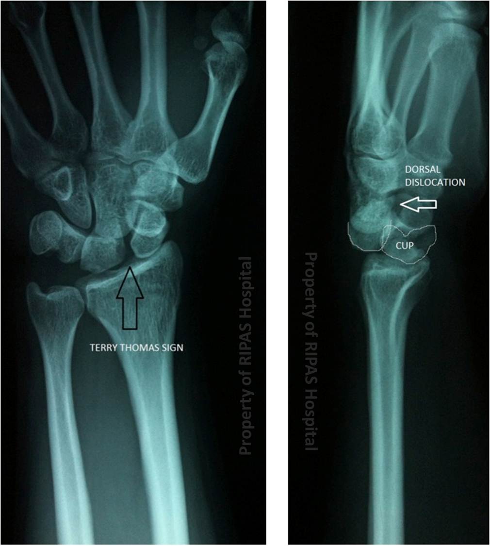
The majority of cases involve dorsal dislocation of the capitate and carpus relative to the lunate which remains in near-normal alignment with the radius. It is always seen best on the lateral wrist film, which is why it should be reviewed with care. The capitate does not sit within the distal articular concavity ('cup') of the lunate, lying dorsal to the lunate.
It can be identified on the AP projection, with widening of the scapholunate space – termed the ‘Terry Thomas sign’ (Figure 2). This is due to its resemblance to the gap between the teeth of the late, famous, comic actor of this name.
|
|
|
Figure 2: Annotated X-ray of the wrist (Click on image to enlarge) |
Perilunate dislocation involves traumatic rupture of the multiple ligaments: radioscaphocapitate, scapholunate interosseous and lunotriquetral interosseous ligaments.
More than half of perilunate dislocations are associated with a trans-scaphoid fracture, which is then termed a trans-scaphoid-perilunate dislocation.
For more details on perilunate dislocation, please click on the following website: http://www.wikiradiography.com/page/Lunate+and+Perilunate+Dislocations
Images and text contributed and prepared by
Dr Ian Bickle, Department of Radiology, RIPAS Hospital.
All images are copyrighted and property of RIPAS Hospital.
![]()