Figure 1a
Figure 1b
IMAGE OF THE WEEK
WEEK 30
|
|
|
|
|
Figure 1a |
Figure 1b |
|
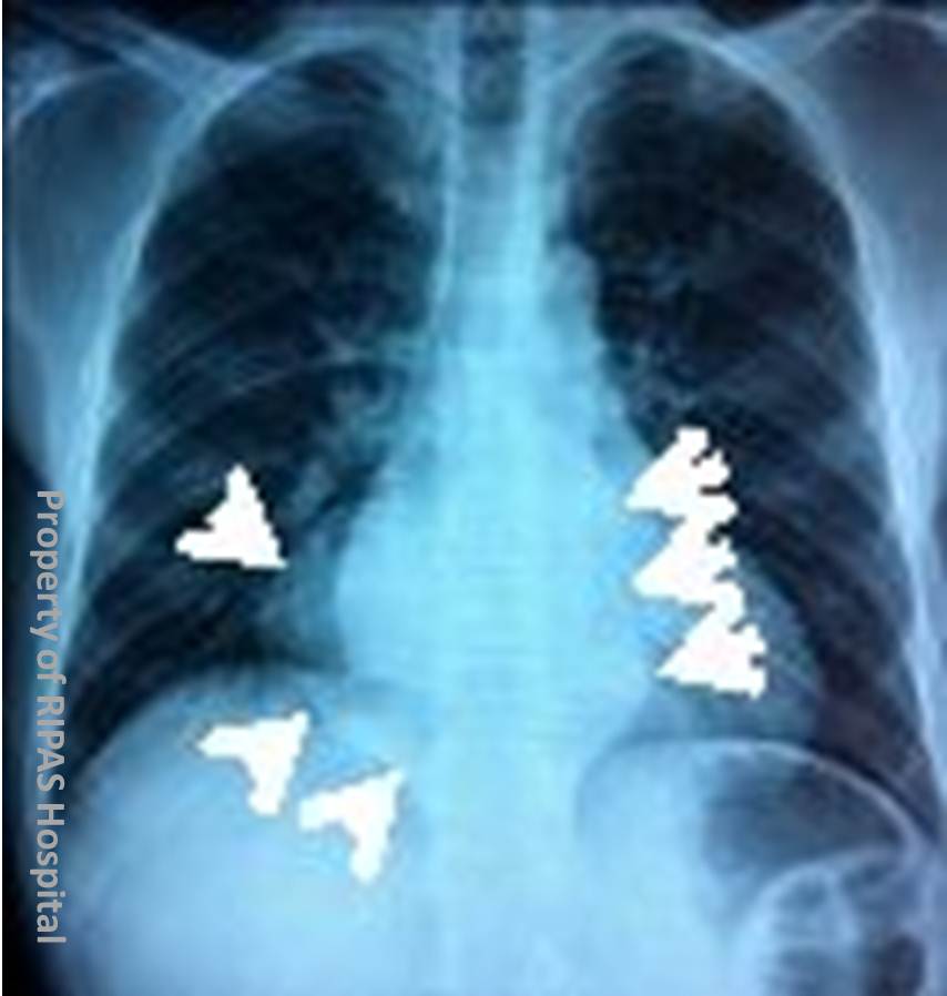
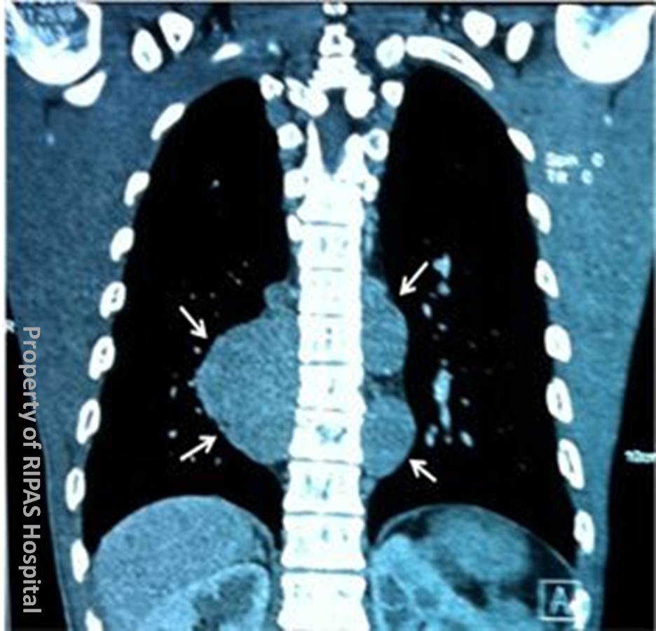
POSTERIOR MEDIASTINAL (PARAVERTEBRAL) MASS - EXTRAMEDULLARY HAEMATOPOIESIS FROM BETA-THALASSAEMIA
The chest x-ray shows a paraspinal mass (posterior mediastinum) in the mid-thoracic region.
Causes of Posterior Mediastinal Mass include:
Primary tumours – in particular neurogenic tumours, such as schwannoma, neurofibroma and neuroblastoma
Metastatic spinal deposits
Infection, in particular a tuberculous paraspinal abscess
Vascular lesions, such as an aortic aneurysm
Paraspinal lymphadenopathy
Congenital lesions, such as enteric or neurogenic cysts
Extramedullary haemopoiesis
Beta-thalassaemia is an inherited, multisystem disorder due to absent or reduced beta-globin chain synthesis resulting in thalassaemia major (homozygotes) or less severe thalassaemia intermedia (heterozygotes). Clinical features include anaemia, hepato-splenomegaly and extramedullary haematopoiesis with secondary skeletal deformity.
The condition is treated with regular blood transfusions, iron chelating agents to prevent iron overload from repeated transfusions, bisphosphonates for co-existing osteoporosis, and splenectomy.
IMAGING
Skeletal
In the skull the following features may be observed:-
Widened diploic space
Coarsened trabeculae and thinned outer table
Frontal bossing
“hair-on-end” appearance, with relative sparing of occipital bone (low marrow content).
Marrow expansion in may occur in the paranasal sinuses with impaired pneumatization of maxillary antra and mastoid sinuses. Marrow hyperplasia in the maxilla causes lateral displacement of the orbits and ventral displacement of the incisors (rodent facies).
Osteopenia
Widened medullary spaces with thinned cortices, premature fusion of epiphyses.
Bulbous widening posterior ribs.
Extra-skeletal
Cardiomegaly, congestive cardiac failure (high output due to anaemia).
Extramedullary haematopoiesis, causing paraspinal masses (arrows on images. Extramedullary haemopoiesis occurs due to a compensatory response to deficient bone marrow blood cell production.
Extramedullary haemopoiesis may occur at an area of foetal erythropoiesis, including the paraspinal regions, adrenal glands, renal pelvis and anterior rib ends.
Heptosplenomegaly
Gallstones.
Iron deposition in organs from repeated transfusions, resulting in high attenuation, appearance of the liver on CT images.
Images contributed by Dr KC Lim, Department of Radiology, RIPAS Hospital, Brunei Darussalam.
All images are copyrighted and property of RIPAS Hospital.
![]()