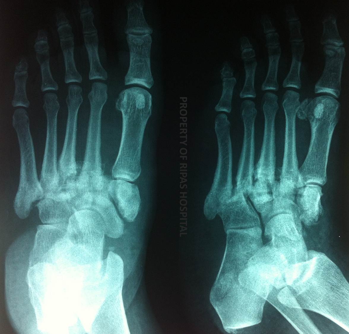
Figure 1: X-ray of the feet showing Charcot's joint deformity.
(Click on image to enlarge)
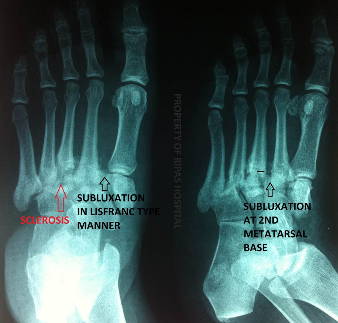
Figure 2: Annotated x-ray of the feet showing Charcot's joint deformity.
(Click on image to enlarge)
IMAGE OF THE WEEK 2012
WEEK 31
Charcot’s Foot
|
|
|
|
Figure 1: X-ray of the feet showing Charcot's joint deformity. (Click on image to enlarge) |
Figure 2: Annotated x-ray of the feet showing Charcot's joint deformity. (Click on image to enlarge) |
A Charcot joint (neuropathic joint) is due to a progressive destructive joint disorder in patients with impaired pain sensation and proprioception. In contemporary medicine, particularly when involving the foot/ankle, this is due to longstanding diabetes.
The 2 theories for the pathophysiology of this condition are the neuro-traumatic theory in which repeated trauma with no sensory feedback occurs and the neurovascular theory in which the absence of neural stimuli results in vasodilatation and hyperemia which promotes bone resorption.
Other less common causes aside from diabetes include the D S’s:
Syphilis
Steriods
Spinal Cord Injury
Syringomyelia
Spina Bifida
Scleroderma
On plain film radiography it is characterised by multiple findings, remembered as the 6 D’s:
Dense bones (sub-chondral sclerosis)
Degeneration
Destruction
Deformity
Debris (loose bodies)
Dislocation
Images and text contributed and prepared by
Dr Ian Bickle, Department of Radiology.
All images are copyrighted and property of RIPAS Hospital.
![]()