IMAGE OF THE WEEK 2012
WEEK 32
OBTURATOR HERNIA
|
 |
 |
|
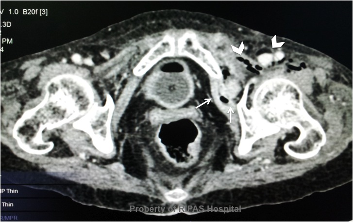
Figure 1: CT pelvis at the hip joint level,
indicating the obturator hernia (white arrows) of small bowel content
passing through the obturator foramen into the thigh with gas formation
in the muscles of the thigh (white arrow heads).
(Click on image to
enlarge) |
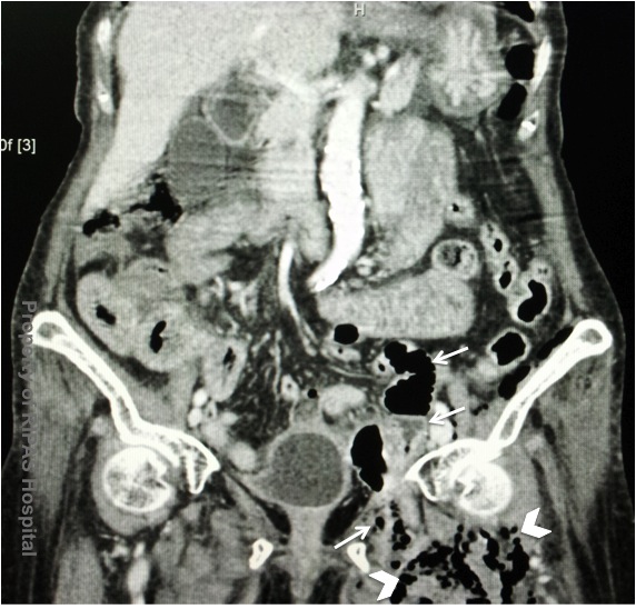
Figure 2: Annotated CT abdomen and pelvis showing
herniation of the small bowel and content through the obturator foramen
(white arrows) and gas formation due to infection and gangrene in the
muscles of the thigh (white arrow heads).
(Click on image to
enlarge) |
An
Obturator Hernia, or pelvic hernia is a rare hernia that occurs when part of the
small bowel passes through the obturator foramen, along the path of the
obturator nerve and muscles. The incidence of obturator hernia is less than 1%
with a female to male ratio of 6:1, because of a gender-specific larger
obturator canal diameter and occurred in predominantly in the (emaciated)
elderly women.
Because
of its anatomic position deep in the pelvis, obturator hernia tends to present
as intestinal obstruction rather than protrusion of intestinal contents.
Patients then to present with a complaint of deep seated hip, thigh or knee pain
and may experience symptoms of loss of appetite, nausea and vomiting with signs
of bowel obstruction such as abdominal distension and abdominal colicky pain.
Diagnosis of a pelvic hernia is often made by eliciting the Howship-Romberg
sign, a pain down the leg when the hip is extended.
Radiological imaging using CT scan as is with the case above is the gold
standard for making a diagnosis of obturator hernia. Emergency laparotomy and
reduction of the incarcerated bowel with excision of infracted bowel is the
treatment of choice in order to safe lives. Delayed presentation and diagnosis
can lead to bowel infarction with gangrene and gas formation in the deep muscles
as indicated by the white arrow heads in the case presented above. Delayed
surgical intervention contributed to a relatively high morbidity and mortality
rate. Once the incarcerated bowel is reduced, the obturator foramen is closed or
repaired using sutures.
Images and text contributed and prepared by
Mr Chee Fui Chong, Department of Surgery.
All
images are copyrighted and property of RIPAS Hospital.


