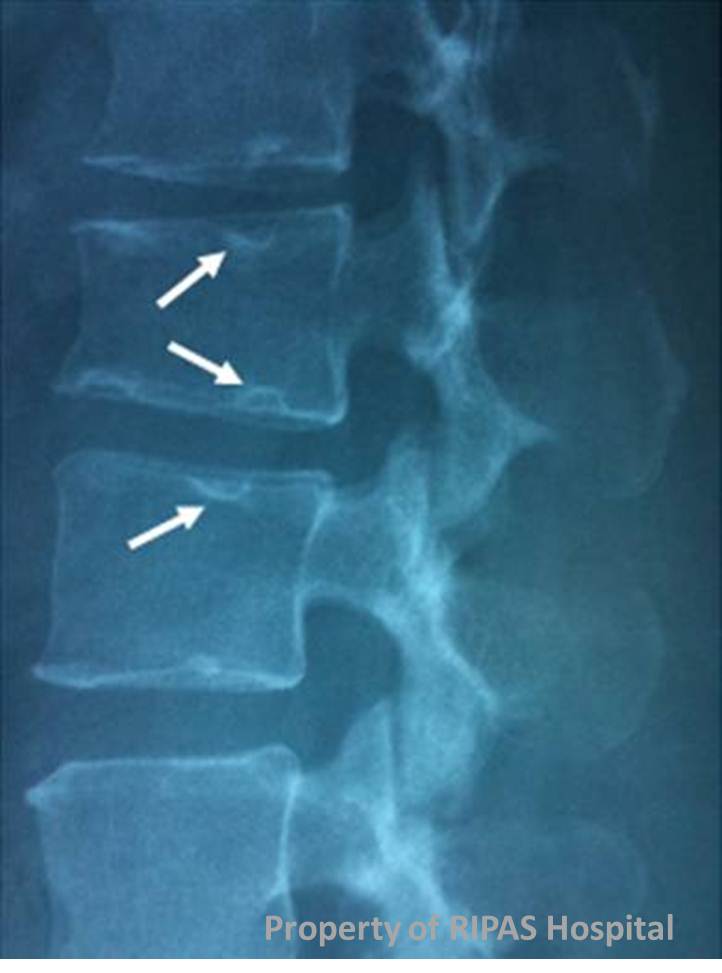
Figure 1: Plain radiograph showing multiple Schmorl’s nodes/nodules.
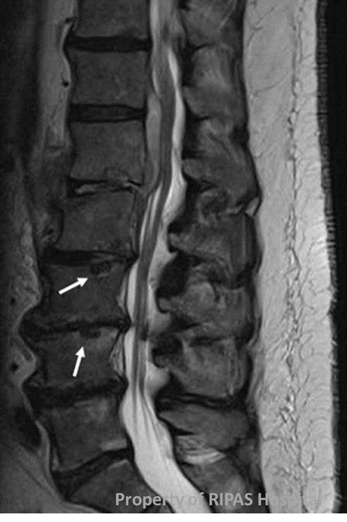
Figure 2: MRI of the same Schmorl's nodes as indicated by white arrows with discitis and epidural abscess at L1-L2 (Refer to Image of the Week, Week 32).
IMAGE OF THE WEEK
WEEK 33
Schmorl’s nodes or nodules
|
|
|
|
|
Figure 1: Plain radiograph showing multiple Schmorl’s nodes/nodules. |
Figure 2: MRI of the same Schmorl's nodes as indicated by white arrows with discitis and epidural abscess at L1-L2 (Refer to Image of the Week, Week 32). |
|
Schmorl’s nodes/nodules are cystic structures on the vertebral plates between the intervertebral cartilages seen on plain radiography of the spine (refer to Figure 1: white arrows). The vertebral bodies most commonly affected are in the middle and lower spine.
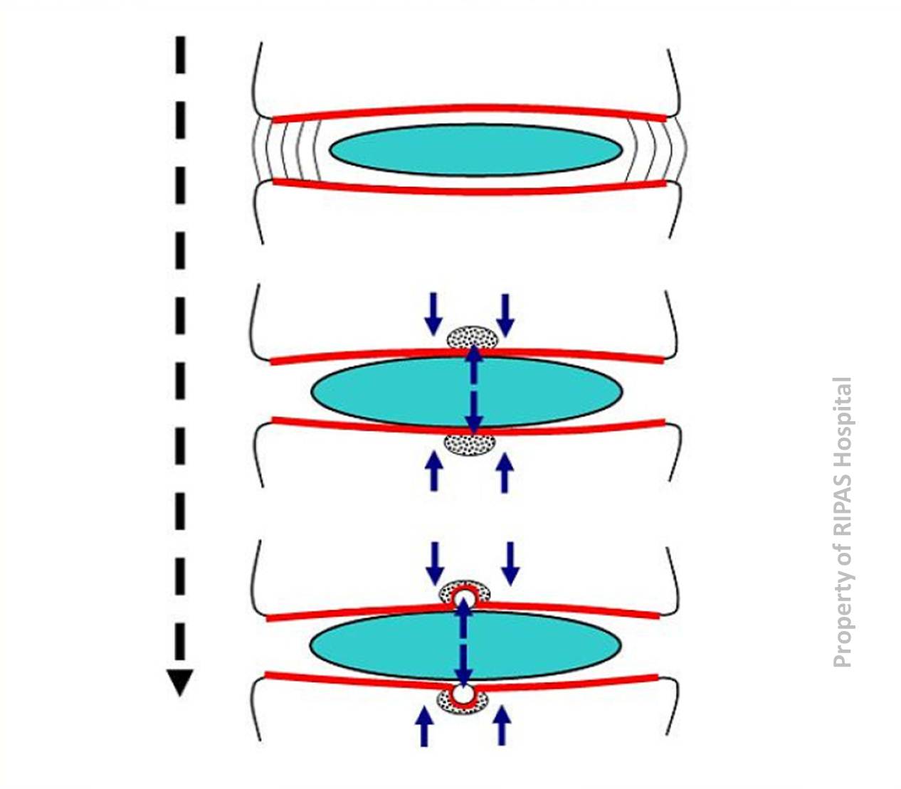
Schmorl’s nodes/nodules are widely believed to be due to upward and downward protrusion (herniation) of the nucleus pulposus of the vertebral discs into the bony tissue of the adjacent vertebrae. Wear and tear of the vertebral discs is thought to weaken the annulus fibrosis resulting in herniation of the nucleus pulposus (Figure 3).
|
|
|
|
Figure 3: Herniation of nucleus pulposus |
Figure 4: Pressure on the end plates results in pressure osteonecrosis with resultant cysts formations. |
However, it has been
shown that protrusions may be secondary rather than the primary events.
Histological examinations show subchondral osteonecrosis and beneath the
cartilagenous endplates, there is fibrosis within the marrow cavities with
disappearance of fat cells and osteocytes. This suggests that ischaemic osteo-necrosis
beneath cartilage endplates with resultant herniation of the nucleus pulposus
and cystic changes (Figure 4).
Schmorl's nodes/nodules are common, especially in the elderly where minor degeneration of the spine and believed to reflect "wear and tear" of the spine. Most patients are asymptomatic and Schmorl’s nodes/nodules are an incidental finding on radiography of the spine. Magnetic resonance imaging (MRI) (Figure 2) is useful and can detect lesions even before they become visible on plain radiography.
Schmorl’s nodes/nodules are named after the German pathologist Christian Georg Schmorl (May 2, 1861 - August 14, 1932) who first described the entity in 1927. Schmorl is best remembered for his work in histology and studies of the human skeleton. He created a histological stain designed to show the canaliculi and lamellae in sections of bone. Among other things, he was also accredited for coining the term (1904) kernicterus to describe nuclear jaundice of the basal ganglia, a condition that was earlier identified by Johannes Orth (pathologist) in 1875.
Images contributed by Dr Chong Vui Heng, Department of Medicine, and Dr Ian Bickle, Department of Radiology, RIPAS Hospital, Brunei Darussalam.
All images are copyrighted and property of RIPAS Hospital.
![]()