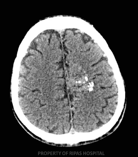
Figure 1
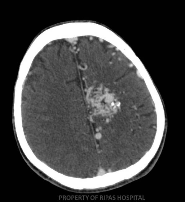
Figure 2
IMAGE OF THE WEEK
WEEK 35
ARTERIOVENOUS MALFORMATION (aVM)
|
|
|
|
|
Figure 1 |
Figure 2 |
|
AVM is a congenital abnormality characterized by a failure of the embryonic vascular plexus to fully differentiate and develop a mature capillary bed. AVM’s consist of a complex network of abnormal vascular channels composed of arterial feeder vessels, arterial collaterals, the AVM nidus, and enlarged draining veins. This results in shunting of blood from the arterial to venous side of the circulation with no intervening intermediary capillary bed. Bleeding is typically from ruptured draining vein.
Presentation is usually, but not exclusively, in early adult life. First presentation is with a seizure (hence why patients presenting with a first seizure should undergo post contrast CT imaging of the brain) or symptoms related to an intracranial haemorrhage. In the non-emergency setting patients may complain of headaches or a progressive neurological defect.
The key figures are:
10% mortality
30% morbidity
2-3% risk of bleeding yearly, increasing to 6% following first bleed and 25% following second bleed.
Imaging Appearances
CT FINDINGS:
NECT: Irregular conglomerate lesion with large feeding arteries and draining veins, and calcified vessels (Figure 1). Acute brain haemorrhage may be seen.
CECT: Strong enhancement of serpiginous dilated vascular bed (Figure 2).
CTA: Enlarged arteries – > nidus – > draining veins.
Images contributed by Dr KC Lim and Dr Ian Bickle, Department of Radiology, RIPAS Hospital, Brunei Darussalam.
All images are copyrighted and property of RIPAS Hospital.
![]()