IMAGE OF THE WEEK
WEEK 4
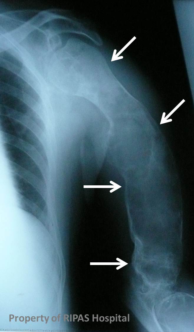
FIBROUS DYSPLASIA
Fibrous dysplasia is a skeletal developmental anomaly of the bone-forming mesenchyme that manifests as a defect in osteoblastic differentiation and maturation. The usual appearance of fibrous dysplasia is a lucent lesion in the diaphysis or metaphysis, with endosteal scalloping. There may or may not be bony expansion. Typically there is no evidence of periosteal reaction, as it is a benign bone lesion. Usually, the matrix of the lesion is smooth and relatively homogeneous; classically described as a ground-glass appearance, where fibrous tissue has replaced the medulla of the bone. Fibrous dysplasia can be either mono or polyostotic, with monostotic accounting for 70-80% and polyostotic for 20-30%. It is not a premalignant condition, but there have been occasional reports of osteosarcoma development. The bone is susceptible to pathological fracture.
McCune-Albright syndrome is polyostotic fibrous dysplasia associated with precocious puberty and cutaneous pigmented lesions
RADIOLOGY
The radiological assessment and classification of bone lesions divides them primarily into aggressive and non-aggressive.
A number of characteristics are taken into account when assessing a bone lesion, which include:
Single or multiple
Location – epiphysis,metaphysis, diaphysis etc
Zone of transition
Expansile or Non-expansile
Lucent or sclerotic
Periosteal reaction
Matrix composition
Images prepared by Dr Ian Bickle, Consultant Radiologist, RIPAS Hospital, Brunei Darussalam.
All images are copyrighted and property of RIPAS Hospital.
![]()