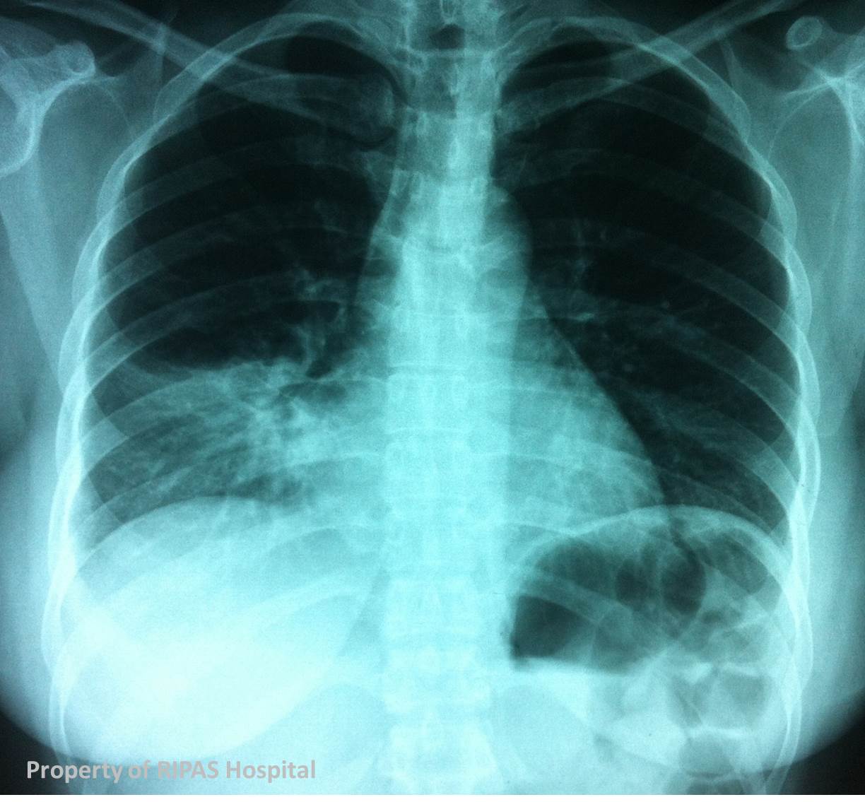
Figure 1a: Normal chest x-ray showing demonstrating the 'Silhouette Sign'.
(Click on image to enlarge)
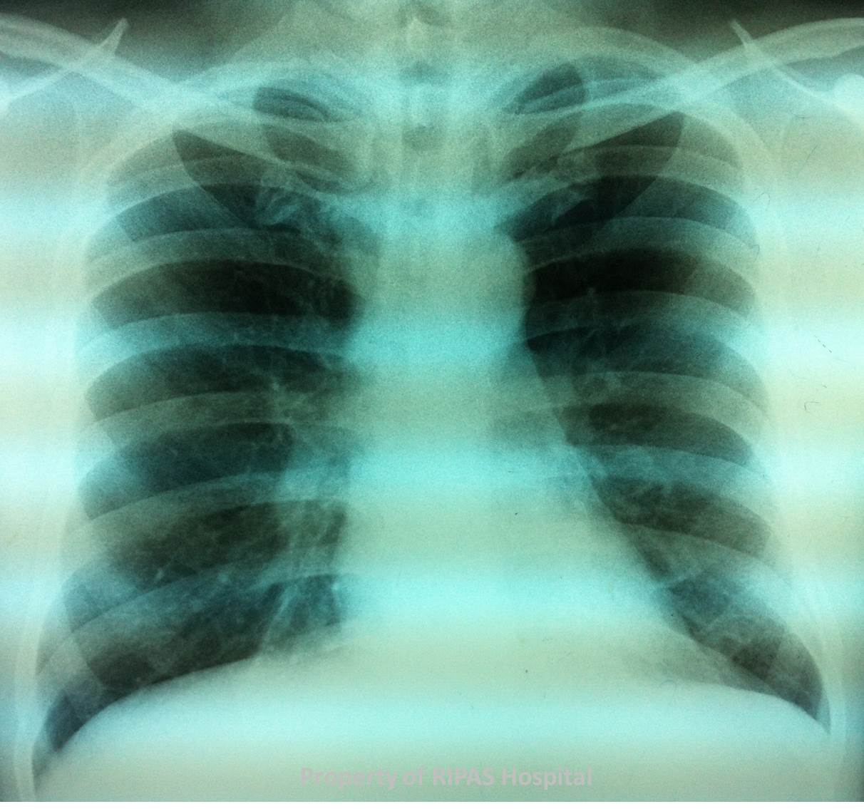
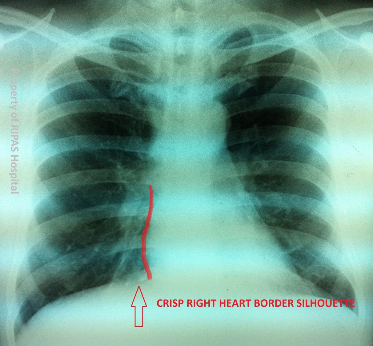
Figure 1b: Annotated normal chest x-ray highlighting (red line) the crisp right heart border silhouette.
(Click on image to enlarge)
IMAGE OF THE WEEK 2013
WEEK 7
The Silhouette Sign
|
|
|
|
Figure 1a: Normal chest x-ray showing demonstrating the 'Silhouette Sign'. (Click on image to enlarge) |
Figure 1b: Annotated normal chest x-ray highlighting (red line) the crisp right heart border silhouette. (Click on image to enlarge) |
One of the greatest radiologists of all time, Benjamin Felson, in his seminal works from the 1950’s coined the term the silhouette sign.
It is a founding and core principle of chest radiograph interpretation – used to identify the lobe in which consolidation or collapse occurs.
On a normal chest radiograph the aerated lung lies adjacent to other structures of different densities (and so different degrees of x-ray absorption occur). This creates a silhouette, which is observed on the chest radiograph.
If the density of the structure is altered – for example the aerated lobe of the lung becomes consolidated or it collapses – its density will increase, it will them absorb more x-rays, and then it will be indistinguishable from the adjacent structure.
For example, a crisp silhouette is observed between the normal right middle lobe and the right heart border, which is formed from the right atrium (Figure 1). When consolidation occurs, like in a pneumonia, the silhouette is lost (Figure 2).
|
|
 |
 |
|
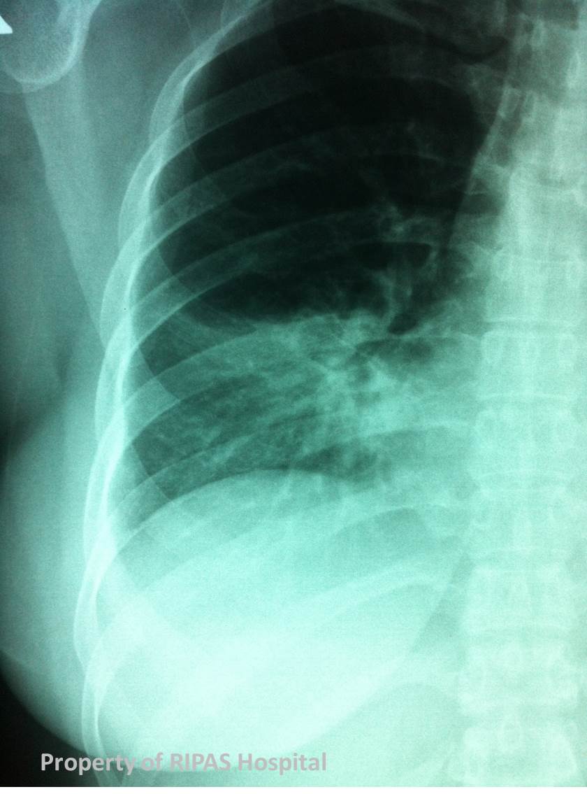
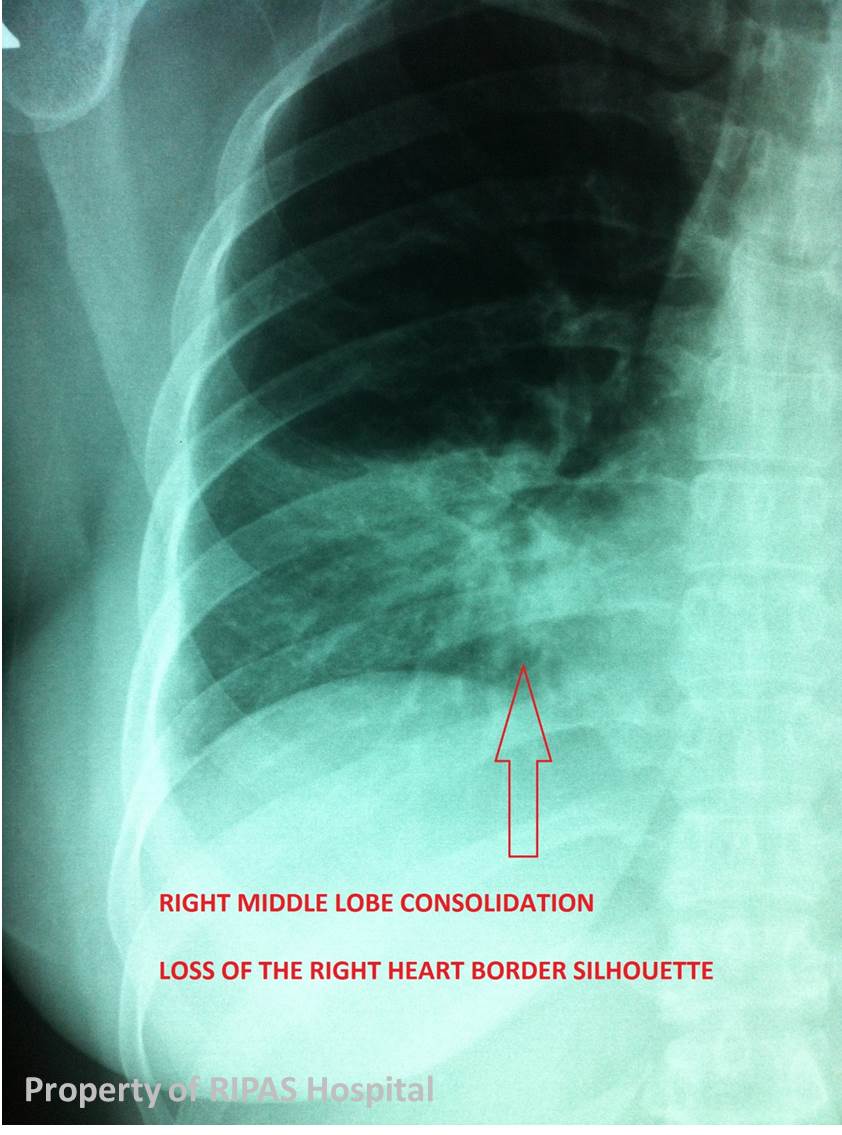
Figure 2a: Chest x-ray of a woman with right middle lobe pneumonia/consolidation, obliterating the normal crisp silhouette sign. (Click on image to enlarge) |
Figure 2b: Close up view of the right middle lobe pneumonia/consolidation with the absence of clear silhouette sign. (Click on image to enlarge) |
Figure 2c: Annotated close up view of the right middle lobe pneumonia/consolidation with the absence of clear silhouette sign . (Click on image to enlarge) |
The same principle applies for all the lobes of the lung.
Left upper lobe: aortic knuckle
Lingula: left heart border
Left lower lobe: left hemidiaphragm
Right upper lobe: right paratracheal stripe
Right middle lobe: right heart border
Right lower lobe: right hemidiaphragm.
Reference
Felson B, Felson H. Localization of intrathoracic lesions by means of the postero-anterior roentgenogram; the silhouette sign. Radiology 1950;55:363-374
Images and text contributed and prepared by
Dr Ian Bickle, Department of Radiology,RIPAS Hospital
All images are copyrighted and property of RIPAS Hospital.
![]()