IMAGE OF THE WEEK
WEEK 7
|
|
|
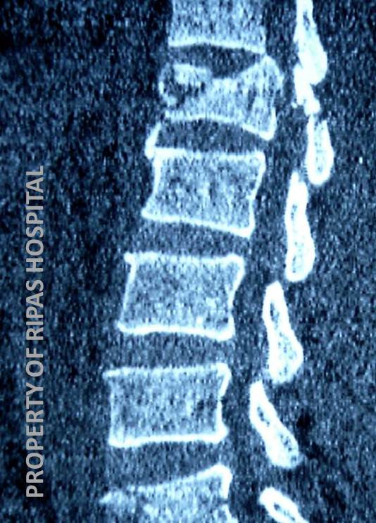
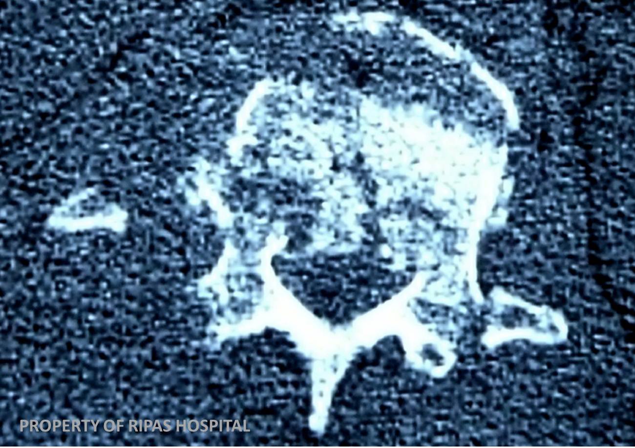
BURST FRACTURE
Typically occurs following a fall from significant height, landing on the feet
(For example, those jumping out of a burning building, off a bridge or from scaffolding).
A burst fracture is a compressive fracture of the vertebral body, with usually both anterior and posterior (retrograde) displacement.
Retrograde displacement can compromise the spinal canal, with or without resultant spinal cord injury.
Can be classified as stable or unstable.
Radiographic Features
On the plain radiographs of the thoracolumbar spine the vertebral bodies of the thoracolumbar junction are typically involved. Assess for loss of height, fracture extent and any retro pulsed fragment(s).
Ensure that appropriate radiographs have been taken to rule out other
concomitant injuries, particularly pelvic and calcaneal fractures.
CT imaging allows assessment of better assessment of the extent of the
injury, any posterior element involvement and spinal canal impingement.
Thos with neurological deficit may also be imaged with MRI to assess injury to the spinal cord.
Images prepared by Dr Ian Bickle, Consultant Radiologist, RIPAS Hospital, Brunei Darussalam.
All images are copyrighted and property of RIPAS Hospital.
![]()