Figure 1: Click on image to enlarge
IMAGE OF THE WEEK 2012
WEEK 9
MENINGIOMA
|
|
|
|
|
Figure 1: Click on image to enlarge |
|
|
Typically a benign intracranial tumour, 90% of which occur in the supra-tentorial brain with a preponderance for the parasagittal/convexity regions (45%).
Usually well visualised on CT, being typically hyperdense on non-contrast images, with intense homogeneous enhancement following IV contrast. The meningioma may contain calcification and cause hyperostosis of the adjacent bone.
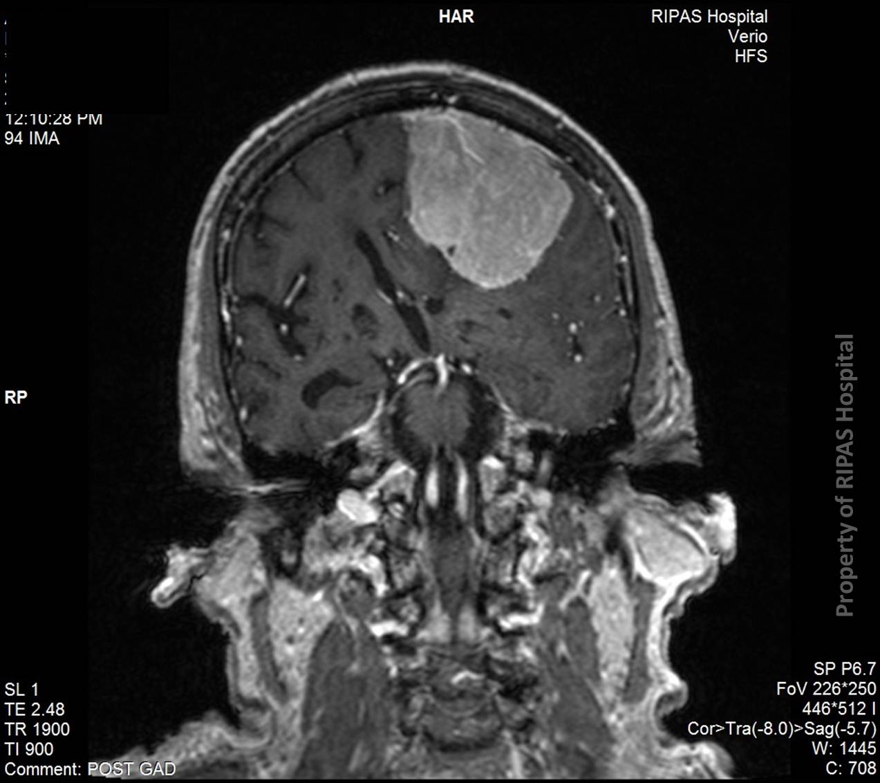
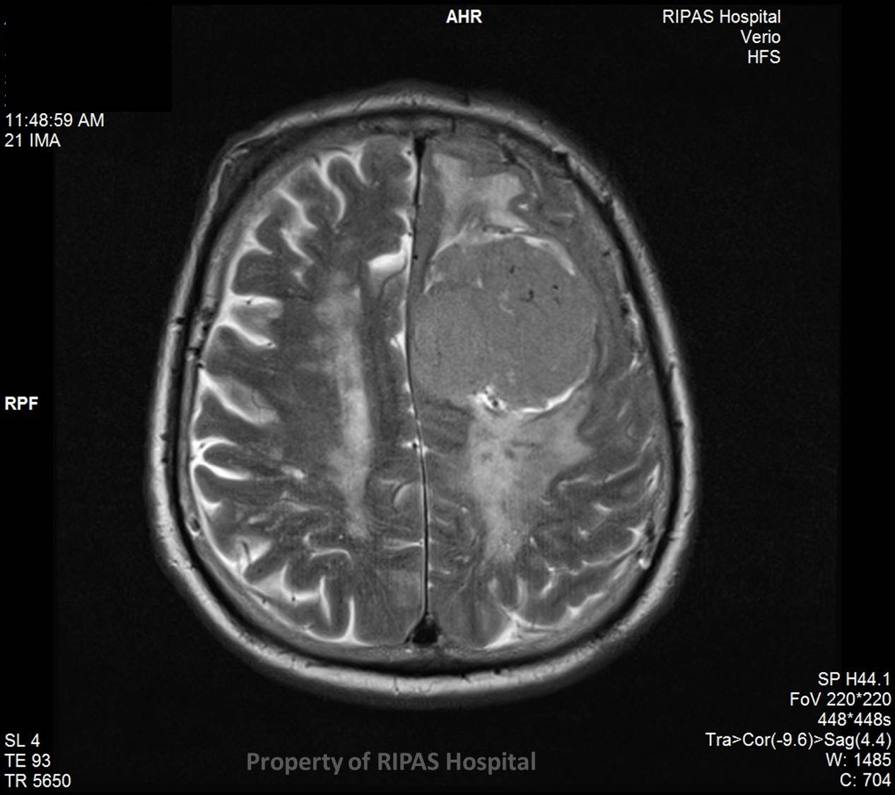
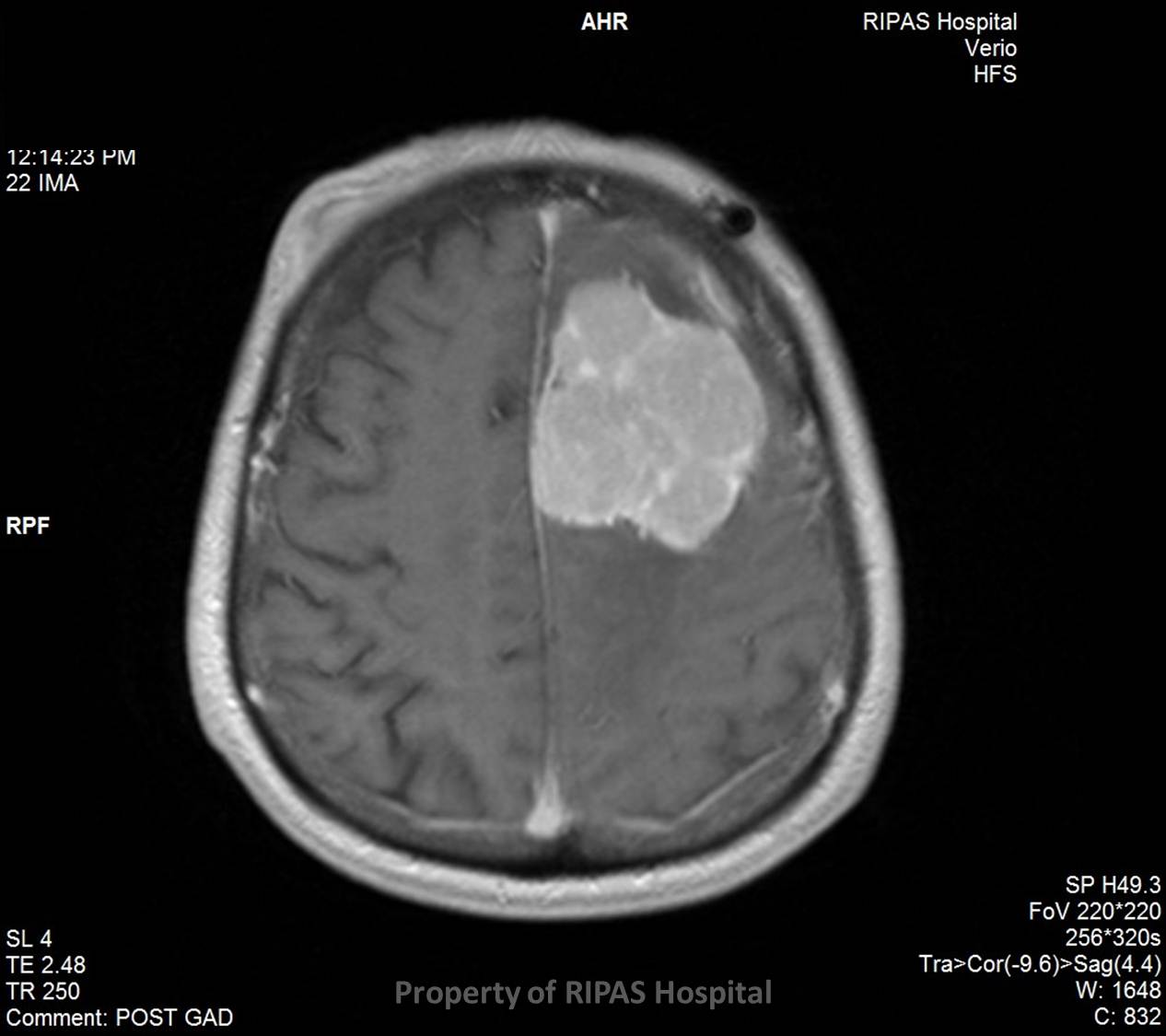
Meningiomas are beautifully illustrated on MRI, the tumour being usually iso/slightly hypointense to the cerebral cortex, with avid homogenous enhancement post contrast (gadolinium) (FigureS 1 & 2). A large proportion demonstrate a ‘dural tail’. There may be a variable amount of peri-tumoral oedema (Figure 3).
Meningiomas are typically solitary. If multiple one must consider the possibility of underlying neurofibromatosis Type 2.
Surgery is typically reserved for those with symptoms, secondary to mass effect from the tumour, or those with features or interval imaging findings raising the rare concern of malignant degeneration.
|
|
|
|
|
Figure 2 |
Figure 3 |
Image and text contributed and prepared by
Dr Ian Bickle, Department of Radiology, RIPAS Hospital, Brunei Darussalam.
All images are copyrighted and property of RIPAS Hospital.
![]()