IMAGE OF THE WEEK
WEEK 9
|
|
|
|
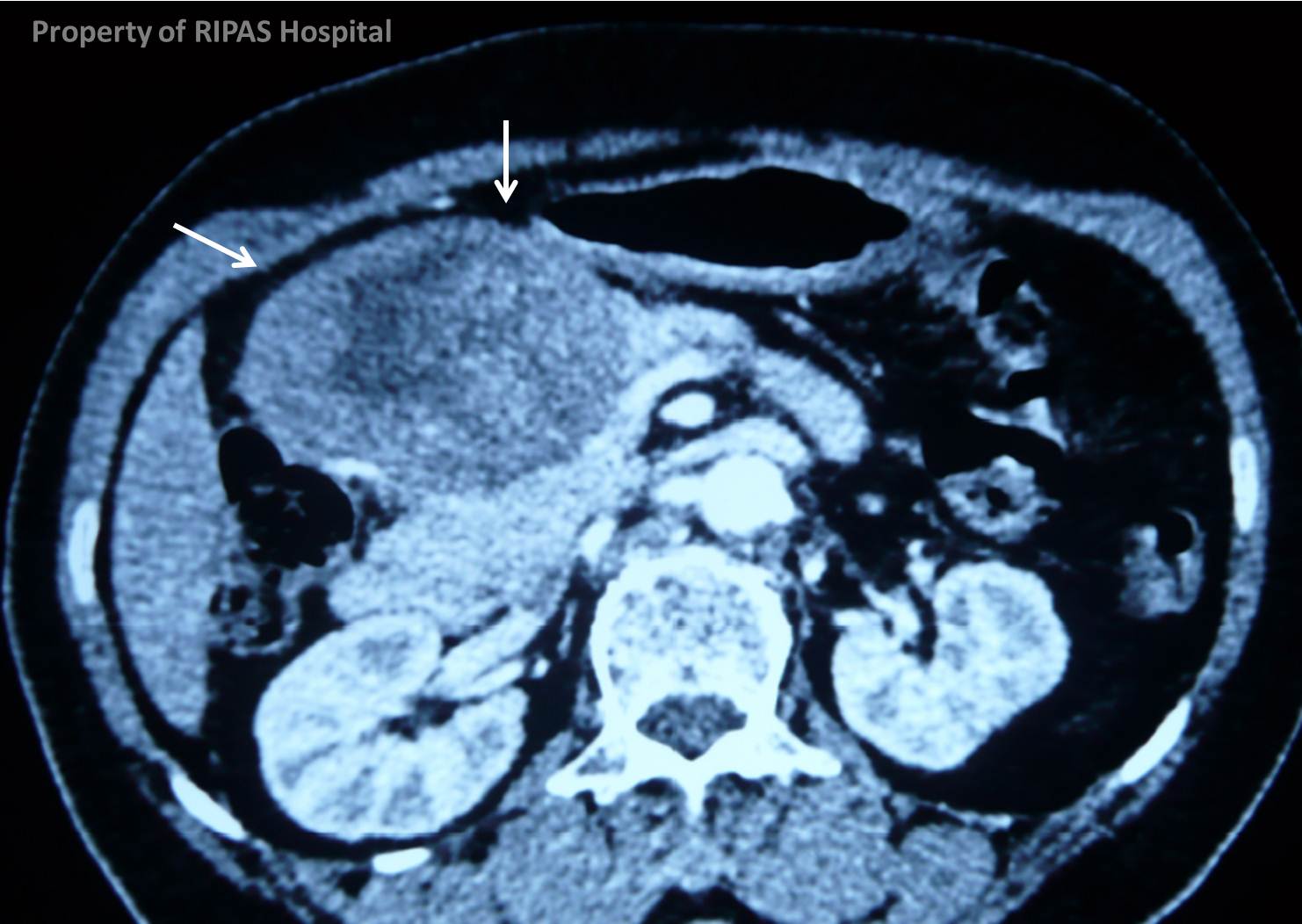
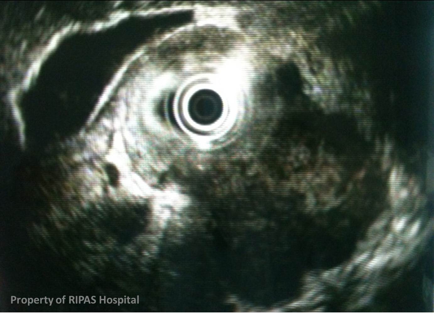
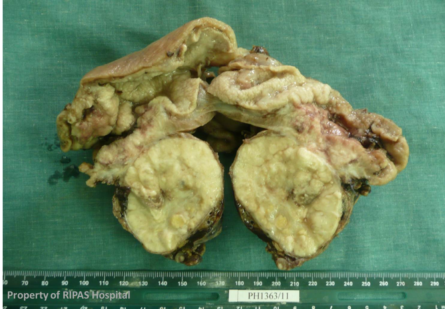
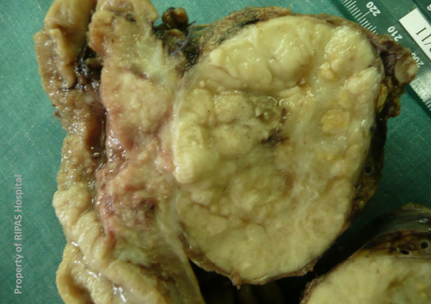
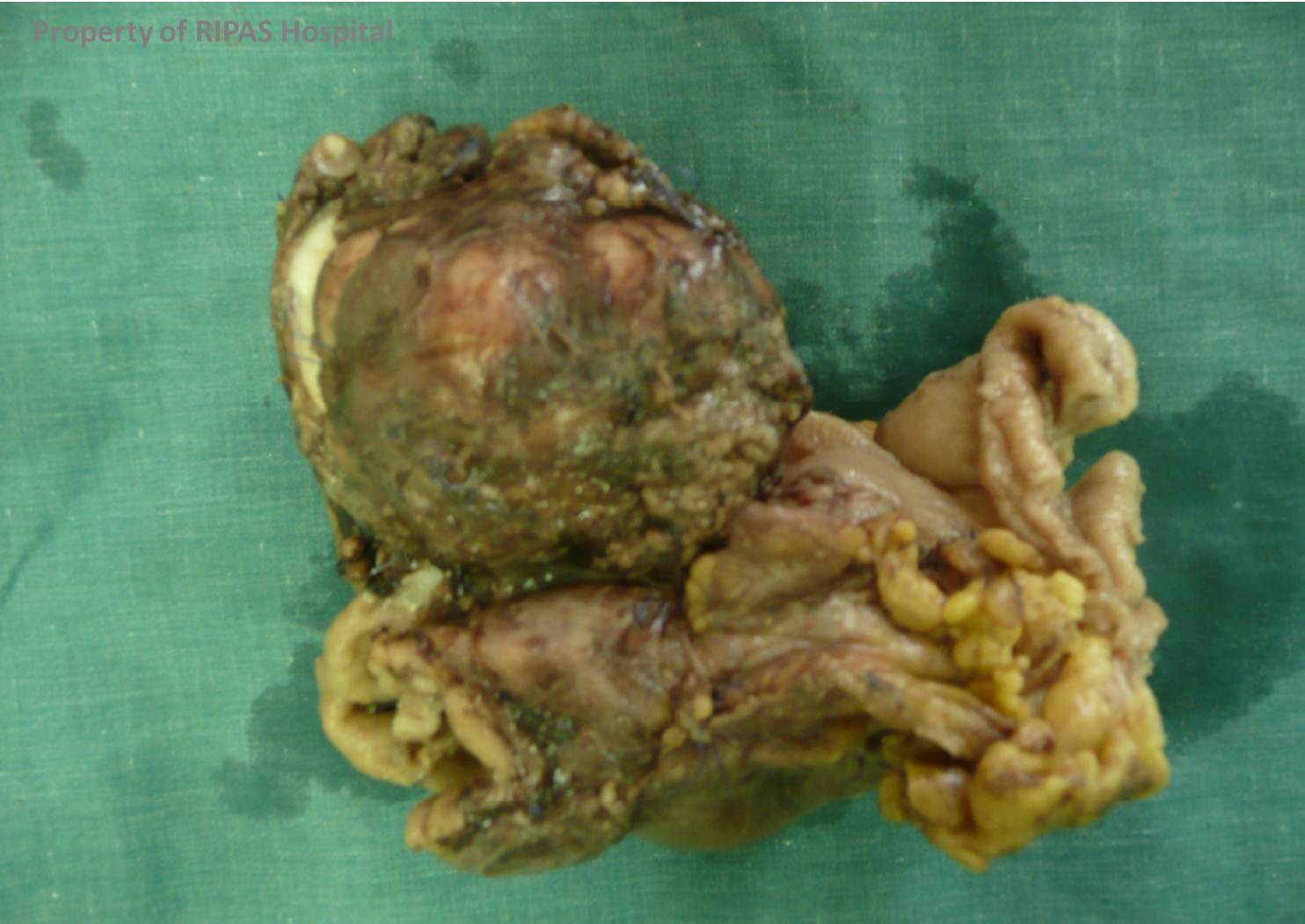
EXOPHYTIC GASTRIC ADENOCARCINOMA
Gastric adenocarcinoma is the most common primary gastrictumour, accounting for approximately 95% of primary gastric malignancies. Gastric adenocarcinoma typically appears as a focal or segmental wall thickening or an intra-luminal mass on CT, providing optimal preparation has been given, such as oral water, to act as a negative contrast agent and to distend the stomach lumen. The gold standard technique for diagnosis is endoscopy with biopsy, not CT, which should be reserved for staging the disease.
As gastric adenocarciomas arise in the mucosa they tend to spread along the gastric wall. Rarely an adenocarcinoma will present as an exophytic gastric mass with a tumour centre lying beyond the confines of the stomach, leading to initial diagnostic difficulties. In particular the mass may mimic a gastrointestinal stromal tumor (GIST). A GIST tends to appear as a well-defined mass that arising from the gastric wall and is commonly exophytic in nature.
Radiographic Features
|
|
|
This collage of images of the CT, endoscopic ultrasound and gross surgical specimens shows the ulcer on the stomach wall, with associated thickening and a large exophytic component.
Learning Point: Exophytic Gastric Adenocarcinoma may mimic a Gastric GIST
Images prepared by Dr Ian Bickle, Consultant Radiologist, RIPAS Hospital, Brunei Darussalam.
All images are copyrighted and property of RIPAS Hospital.
![]()