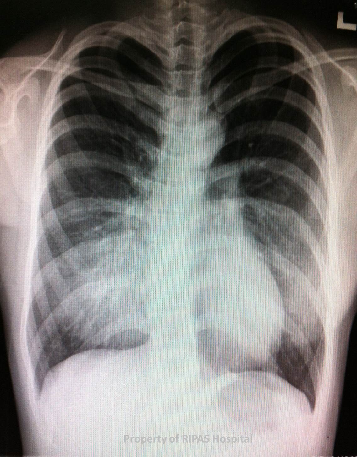
Figure 1a: A plain chest radiograph of a patient with Pectus Excavatum
(Click on image to enlarge)
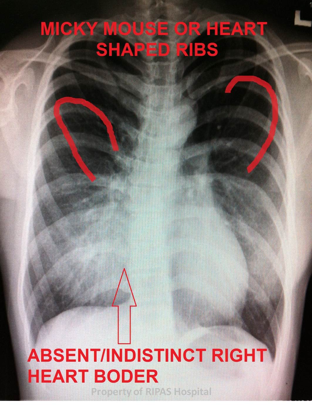
Figure 1b: Annotated plain chest radiograph of a patient with Pectus Excavatum, indicating the unique features seen.
(Click on image to enlarge)
IMAGE OF THE WEEK 2013
WEEK 14
PECTUS EXCAVATUM: PLAIN CHEST RADIOGRAPH FEATURES
|
|
|
|
|
Figure 1a: A plain chest radiograph of a patient with Pectus Excavatum (Click on image to enlarge) |
Figure 1b: Annotated plain chest radiograph of a patient with Pectus Excavatum, indicating the unique features seen. (Click on image to enlarge) |
|
Image of the week featured Pectus Excavatum early last year (Week 3(15-1-2012)). This week Image of Week details the typical plain chest radiographs features associated with patients with Pectus Excavatum as seen in figure 1b, namely:
· Blurring or indistinct right heart border on PA or AP film
· Shift of cardiomediastinal outline to the left
· Crowding of ribs with a horizontal posterior course and a more vertical anterior course than the normal, appearing like heart shaped or Mickey Mouse’s ears
· Increase density of the inframedial lung zone
· Obliteration of the descending thoracic aorta interface
Please refer to Week 3(15-1-2012) for further clinical features and management of Pectus Excavatum
Images contributed by
Dr Ian Bickle, Department of Radiology,RIPAS Hospital
Text prepared by
Dr Chong Chee Fui, Department of Surgery, RIPAS Hospital
All images are copyrighted and property of RIPAS Hospital.
![]()